. 2017 Mar 30;7(1):509.
doi: 10.1038/s41598-017-00626-6.

Interplay of DNA methyltransferase 1 and EZH2 through inactivation of Stat3 contributes to β-elemene-inhibited growth of nasopharyngeal carcinoma cells

Affiliations

Interplay of DNA methyltransferase 1 and EZH2 through inactivation of Stat3 contributes to β-elemene-inhibited growth of nasopharyngeal carcinoma cells

JingJing Wu et al. Sci Rep. .

Abstract

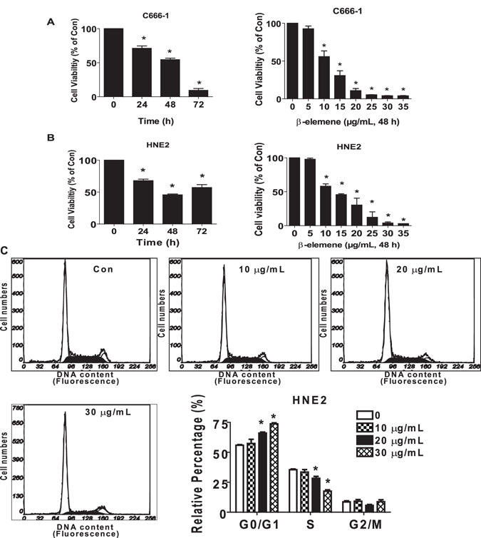
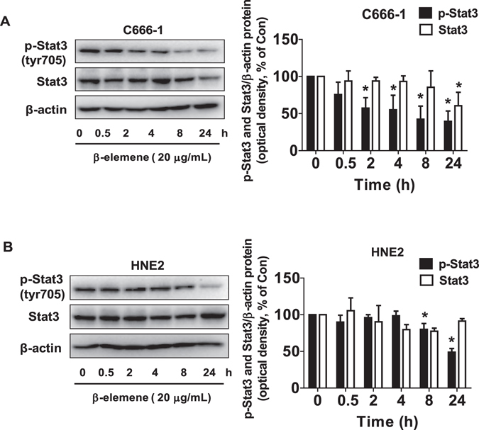
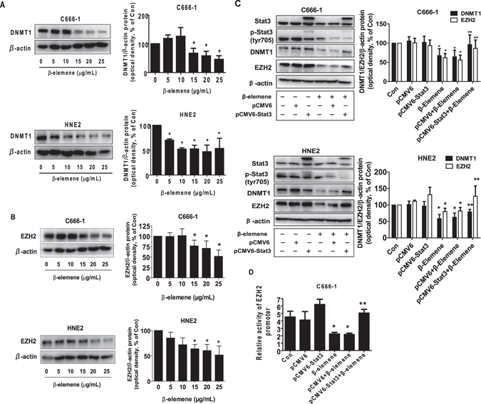
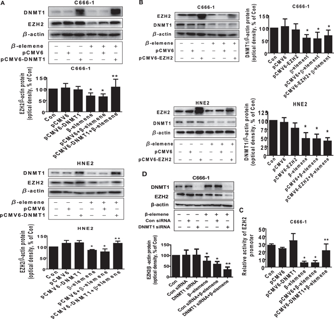
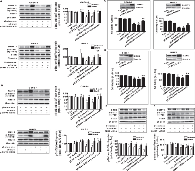
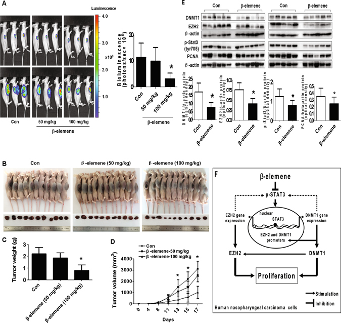
β-elemene, a compound extracted from Curcuma wenyujin plant, exhibits anticancer activity in many cancer types. However, the detailed mechanism by which β-elemene inhibits growth of nasopharyngeal carcinoma (NPC) cells remains unknown. We showed that β-elemene reduced phosphorylation of signal transducer and activator of transcription 3 (Stat3), and protein expressions of DNA methyltransferase 1 (DNMT1) and enhancer of zeste homolog 2 (EZH2). Exogenously expressed Stat3 antagonized the effect of β-elemene on DNMT1 and EZH2 expressions. Furthermore, overexpressions of DNMT1 and EZH2 reversed the effect of β-elemene on phosphorylation of Stat3 and cell growth inhibition. Intriguingly, exogenously expressed DNMT1 overcame β-elemene-inhibited EZH2 protein expression and promoter activity. On the contrary, silencing of EZH2 and DNMT1 genes feedback strengthened the effect of β-elemene on phosphorylation of Stat3. Consistent with this, β-elemene inhibited tumor growth, phosphorylation of Stat3, expressions of DNMT1 and EZH2 in a mouse xenograft model. Collectively, this study shows that β-elemene inhibits NPC cell growth via inactivation of Stat3, and reduces DNMT1 and EZH2 expressions. The interplay of DNMT1 and EZH2, and the mutual regulations among Stat3, EZH2 and DNMT1 contribute to the overall responses of β-elemene. This study uncovers a novel mechanism by which β-elemene inhibits growth of NPC cells.


Conflict of interest statement

The authors declare that they have no competing interests.

Figures

Figure 1
Figure 1
β-elemene inhibited growth and induced cell cycle arrest in NPC cells. (A,B) C666-1 (A) and HNE2 (B) cells were treated with increased concentrations of β-elemene for up to 48 h or to β-elemene (20 μg/mL) for up to 72 h. (C) HNE2 cells were treated with indicated concentrations of β-elemene for 24 h. The cells were collected and processed for analysis of cell cycle distribution. Cell cycle was analyzed by flow cytometry after propidium iodide (PI) staining, and the percentages of the cell population in each phase (G0/G1, S and G2/M) of cell cycle were analyzed by Multicycle AV DNA Analysis Software. Data are expressed as a percentage of total cells. Values are given as the mean ± SD, from 3 independent experiments performed in triplicate. *Indicates significant difference as compared to the untreated control group (P < 0.05).
Figure 2
Figure 2
β-elemene reduced the phosphorylation of Stat3. (A,B) C666-1 (A) and HNE2 (B) cells were treated with β-elemene (20 μg/mL) in the indicated times, and cell lysate was harvested and the expression of the phosphorylated or total protein of Stat3 was measured by Western blot analysis using corresponding antibodies. β-actin was used as loading control. The figures are representative cropped gels/blots that have been run under the same experimental conditions. The bar graphs represented the densitometry results of p-Stat3, Stat3/β-actin as mean ± SD of at least three separate experiments. *Indicates significant difference from the untreated control cells (P < 0.05).
Figure 3
Figure 3
β-elemene inhibited the protein expression of DNMT1 and EZH2 through Stat3. (A,B) C666-1 and HNE2 cells were exposed to increased concentrations of β-elemene for 24 h, followed by measuring DNMT1 and EZH2 proteins by Western blot. (C) C666-1 and HNE2 cells were transfected with control or Stat3 expression vector described in the Materials and Methods section for 24 h followed by exposure the cells to β-elemene (20 μg/mL) for an additional 24 h. Afterwards, Stat3, p-Stat3, DNMT1 and EZH2 protein expressions were determined by Western blot. (D) C666-1 cells were transfected with control or Stat3 expression vector, and with a wild type human EZH2 promoter reporter construct ligated to luciferase reporter gene and internal control secreted alkaline phosphatase for 24 h, followed by treating with β-elemene (20 μg/mL) for an additional 24 h. Afterwards, the promoter activities were determined using the Secrete-Pair Dual Luminescence Assay Kit as described in the Materials and Methods section. Values in bar graphs were given as the mean ± SD from three independent experiments performed in triplicate. *Indicates significant difference as compared to the untreated control group (P < 0.05). **Indicates significant difference from the β-elemene treated alone (P < 0.05).
Figure 4
Figure 4
Exogenously expression of DNMT1 overcame the effect of β-elemene on EZH2 protein expression and promoter activity. (A,B) C666-1 and HNE2 cells were transfected with control or DNMT1 or EZH2  expression vectors for 24 h prior to exposure of the cells to β-elemene (20 μg/mL) for an additional 24 h. Afterwards, Western blot analysis were used measure the protein levels of DNMT1 and EZH2 using corresponding antibodies. (C) C666-1 and HNE2 cells were transfected with control or DNMT1 expression vector, and with a wild type human EZH2 promoter reporter construct ligated to luciferase reporter gene and internal control secreted alkaline phosphatase for 24 h, followed by treating with β-elemene (20 μg/mL) for an additional 24 h. Afterwards, the promoter activities were determined using the Secrete-Pair Dual Luminescence Assay Kit as described in the Materials and Methods section. (D) C666-1 cells were transfected with control or DNMT1 siRNA for 24 h prior to exposure of the cells to β-elemene (20 μg/mL) for an additional 24 h. Afterwards, Western blot analysis were used for determining the protein levels of DNMT1 and EZH2 using corresponding antibodies. The figures are representative cropped gels/blots that have been run under the same experimental conditions. Values in bar graphs were given as the mean ± SD from three independent experiments performed in triplicate. *Indicates significant difference as compared to the untreated control group (P < 0.05). **Indicates significant difference from the β-elemene treated alone (P < 0.05).
Figure 5
Figure 5
Exogenously expression of DNMT1 or EZH2 feedback reversed the effect of β-elemene on phosphorylation of Stat3 and cell growth inhibition. (A,B) C666-1 and HNE2 cells were transfected with control or DNMT1 or EZH2 expression vectors for 24 h prior to exposure of the cells to β-elemene (20 μg/mL) for an additional 24 h. Afterwards, Western blot analysis were used measure the levels of DNMT1 and EZH2, and p-Stat3, Stat3 proteins using corresponding antibodies. (C,D) C666-1 and HNE2 cells were transfected with control or DNMT1 or EZH2 expression vectors for 24 h prior to exposure of the cells to β-elemene (20 μg/mL) for an additional 48 h. Afterwards, cell proliferation was examined by MTT assays as described in the Materials and Methods section. Insert blots represented protein expressions of DNMT1 and EZH2. (E,F) C666-1 cells were transfected with control or DNMT1 or EZH2 siRNAs for 30 h prior to exposure of the cells to β-elemene (20 μg/mL) for an additional 24 h. Afterwards, Western blot analysis were used measure the levels of DNMT1 and EZH2, and p-Stat3, Stat3 protein using corresponding antibodies. The figures are representative cropped gels/blots that have been run under the same experimental conditions. Values in bar graphs were given as the mean ± SD from three independent experiments performed in triplicate. *Indicates significant difference as compared to the untreated control group (P < 0.05). **Indicates significant difference from the β-elemene treated alone (P < 0.05).
Figure 6
Figure 6
In vivo anti-tumor efficacy of β-elemene in subcutaneous lung tumor-bearing nude mice model. Mice (n = 11/group) were divided to 3 groups [Con (saline), Low (L, 50 mg/kg) and High (H, 100 mg/kg)], and β-elemene was given at the 10th day after tumor cells injection by gavages for up to 17 days. (A) The xenografts were assessed by in vivo bioluminescence imaging at the day 1 and the end of the experiments (on day 17). The tumor growth was monitored by injecting luciferin in the mice followed by measuring bioluminescence using IVIS Imaging System. Imaging and quantification of signals were controlled by the acquisition and analysis software living image as described in the Materials and Methods section. Representative images are shown. (B) The photographs of β-elemene or vehicle-treated xenografts derived from nude mice are shown. (C,D) The xenografts were harvested on day 17, and the volume and weight of tumors were measured. (E) At the end of the experiments, xenografted tumors from the high dose and control groups were isolated from individual animals and the corresponding lysates were processed for detecting DNMT1, EZH2, p-Stat3, and PCNA by Western blot. β-actin was used as loading control. The bar graphs represented the tumor weight and volume of mice results of as mean ± SD. *Indicates the significant difference from the untreated control (p < 0.05). (F) The diagram shows that β-elemene inhibits NPC growth through inactivation of Stat3, followed by reduction of DNMT1 and EZH2 expression. The reciprocal communication between DNMT1 and EZH2, the mutual regulatory loops of Stat3 with DNMT1 and EZH2 contribute to the overall responses of β-elemene in this process.

Similar articles

Cited by

References

    1. Pan Y, Claret FX. Targeting Jab1/CSN5 in nasopharyngeal carcinoma. Cancer Lett. 2012;326:155–160. doi: 10.1016/j.canlet.2012.07.033. - DOI - PMC - PubMed
    1. Hildesheim A, Wang CP. Genetic predisposition factors and nasopharyngeal carcinoma risk: a review of epidemiological association studies, 2000-2011: Rosetta Stone for NPC: genetics, viral infection, and other environmental factors. Semin Cancer Biol. 2012;22:107–116. doi: 10.1016/j.semcancer.2012.01.007. - DOI - PMC - PubMed
    1. Yang G, et al. The inhibitory effects of extracellular ATP on the growth of nasopharyngeal carcinoma cells via P2Y2 receptor and osteopontin. J Exp Clin Cancer Res. 2014;33:53. doi: 10.1186/1756-9966-33-53. - DOI - PMC - PubMed
    1. Bruce JP, Yip K, Bratman SV, Ito E, Liu FF. Nasopharyngeal Cancer: Molecular Landscape. J Clin Oncol. 2015;33:3346–3355. doi: 10.1200/JCO.2015.60.7846. - DOI - PubMed
    1. Wang S, Zhang R, Claret FX, Yang H. Involvement of microRNA-24 and DNA methylation in resistance of nasopharyngeal carcinoma to ionizing radiation. Mol Cancer Ther. 2014;13:3163–3174. doi: 10.1158/1535-7163.MCT-14-0317. - DOI - PMC - PubMed

Publication types

MeSH terms

LinkOut - more resources