. 2017 Mar 30;7(1):512.
doi: 10.1038/s41598-017-00658-y.

Chromosomal integration of HHV-6A during non-productive viral infection

Affiliations

Chromosomal integration of HHV-6A during non-productive viral infection

Nitish Gulve et al. Sci Rep. .

Abstract

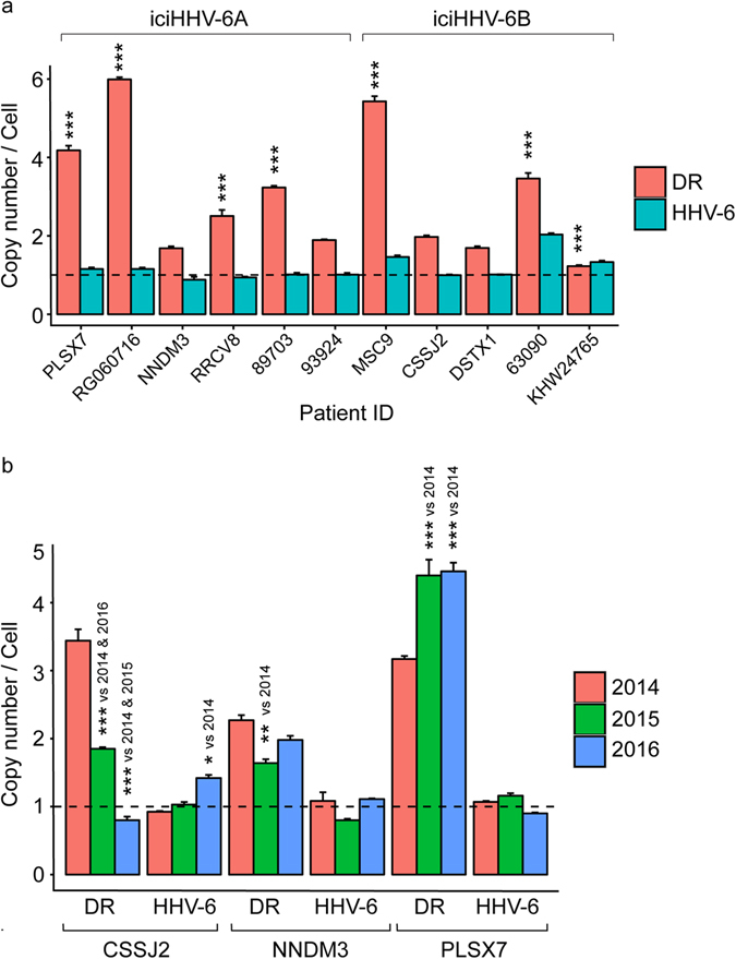
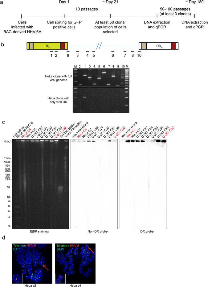
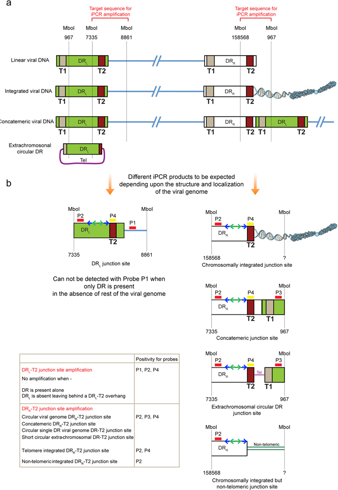
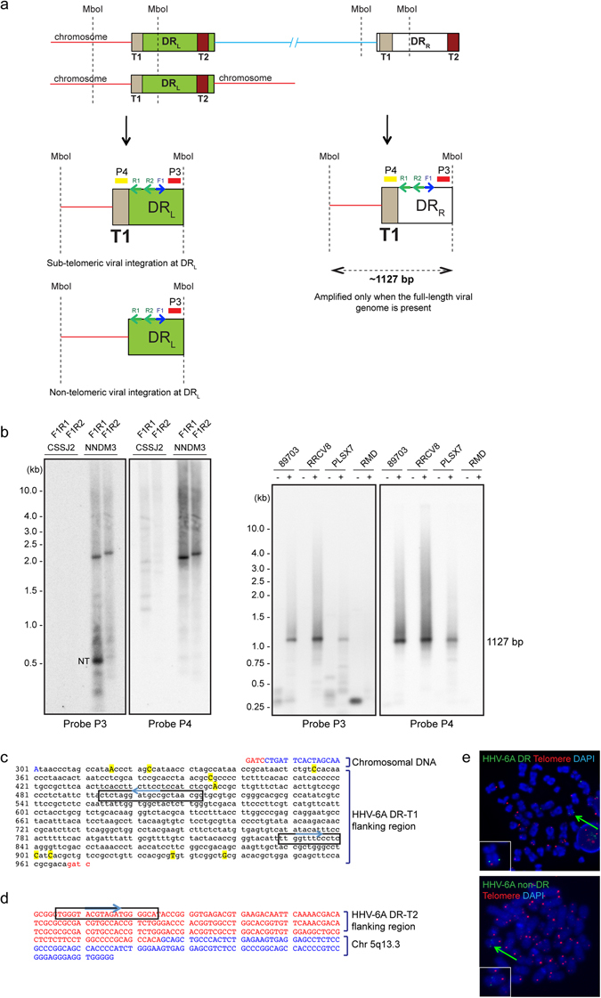
Human herpesvirus 6A (HHV-6A) and 6B (HHV-6B) are two different species of betaherpesviruses that integrate into sub-telomeric ends of human chromosomes, for which different prevalence rates of integration have been reported. It has been demonstrated that integrated viral genome is stable and is fully retained. However, study of chromosomally integrated viral genome in individuals carrying inherited HHV-6 (iciHHV-6) showed unexpected number of viral DR copies. Hence, we created an in vitro infection model and studied retention of full or partial viral genome over a period of time. We observed an exceptional event where cells retained viral direct repeats (DRs) alone in the absence of the full viral genome. Finally, we found evidence for non-telomeric integration of HHV-6A DR in both cultured cells and in an iciHHV-6 individual. Our results shed light on several novel features of HHV-6A chromosomal integration and provide valuable information for future screening techniques.


Conflict of interest statement

The authors declare that they have no competing interests.

Figures

Figure 1
Figure 1
High number viral DRs are detected in many iciHHV-6 individuals. (a) Number of copies of HHV-6 genome and viral DR per cell were quantified using qPCR. HHV-6 and DR copy numbers for different individuals were compared against patient 93924 for statistical analysis as 93924 had ~1 copy of HHV-6A and ~2 copies of DR representing the most ideal scenario. (b) Viral genome copy numbers and DR copy numbers were quantified over a period of 3 years in 3 different iciHHV-6 individuals. Data represents mean values of 3 different biological triplicates. Statistical analysis was done by using two-way Analysis of Variance (ANOVA) followed by Tukey’s Honest Significance Differences (HSD) test for determination of individual comparisons, adjusted P values. * < 0.05, ** < 0.01, *** < 0.001. Copy number of 1 is marked as a baseline (dotted line).
Figure 2
Figure 2
In vitro cell culture infection model to study chromosomal integration of HHV-6. (a) Diagrammatic representation of the experimental set up. (b) 10 different regions of HHV-6A genome in 2 different clones of HeLa cells were tested by conventional PCR. Upper panel represent approximate location of primer pairs within HHV-6A genome. Lower panel shows ethidium bromide stained agarose gel images showing amplified PCR products. M, 1 kb DNA ladder; 1, DR6; 2, DR7; 3, U22; 4, P41; 5, U42; 6, U79; 7, U83; 8, U91; 9, DRL junction region; 10, DRR junction region. (c) Presence of chromosome associated full-length viral genome and viral DR was studied in various HeLa and U-251 clones by PFGE. Blots were first hybridised with a probe that does not detect HHV-6 DR (non-DR probe), stripped and then re-probed with a DR specific probe (DR probe). Cells positive for viral DR alone are indicated in red. (d) FISH analysis of integrated HHV-6A in HeLa cells. Left panel shows integrated viral genome in clone 2. Co-staining with telomere specific probe is indicated. The right panel shows the integration of viral DR in clone 4. HHV-6A DR specific probe is used for viral genome detection. Arrowhead indicates integrated viral genome, which is also shown as enlarged images on lower left hand corner of each image.
Figure 3
Figure 3
Schematic representation of the inverse PCR experimental set up to amplify DR-T2 junction region. (a) Various conformations of HHV-6 genome reported so far are compared on the backdrop of the inverse PCR. MboI is a frequent cutter restriction enzyme. Hence, only the cut sites of our interest are indicated. Nucleotide position of the MboI cut sites (reference genome sequence X83413.1) is indicated. DRL, left direct repeat; DRR, right direct repeat; T1, heterogeneous telomeric repeat sequence; T2, homogeneous telomeric repeat sequence. (b) Different combinations of iPCR products expected are described depending upon the structure and integration status of the viral genome. Locations of the 4 hybridisation probes (P1-P4) are marked. First sets of inverse primers are indicated with blue arrows and nested primer pairs are indicated with green arrows. A combination of probes that can detect specific viral structures is indicated within a Table.
Figure 4
Figure 4
Non-telomeric chromosomal integration of HHV-6A. (a) iPCR products from three different cells were checked by Southern hybridisation using 4 different probes as explained in Fig. 3. Potential bands that did not hybridise with a telomere probe (Probe P4) are marked with red arrowhead and were processed for sequence identification. (be) Sequencing and identification of non-telomeric junction site of HHV-6A in four different clonal populations of cells. (b) Sequence analysis of junction site of HHV-6A in SK-OV-3 clone 33. (c) Sequence analysis of junction site of HHV-6A in HeLa clone 2. (d) Sequence analysis of junction site of HHV-6A in HeLa clone 4. (e) Sequence analysis of junction site of HHV-6A in U-251 clone 15. Viral DNA sequences are marked in black whereas chromosomal DNA sequences are marked in blue.
Figure 5
Figure 5
iPCR identified non-telomeric chromosomal integration of HHV-6A in iciHHV-6 individuals. (a) Schematic representation of iPCR to amplify DR-T1 junction site. Different combinations of iPCR products expected are described depending upon the structure and integration status of the viral genome. Locations of the 2 hybridisation probes (P3 and P4) are marked. F1, forward primer; R1 and R2, two reverse primers. (b) iPCR products from 4 different iciHHV-6A individuals (NNDM3, PLSX7, RRCV8 and 89703), a iciHHV-6B individual (CSSJ2) and non-HHV-6 control (RMD) were analysed by Southern hybridisation using two different probes. ~1.1 kb amplimer is expected from a full-length viral genome irrespective of integration status. CSSJ2 was used as a negative control to check primer specificity. NT, possible non-telomeric integration. (c) Sequence analysis of non-telomeric junction site at HHV-6A DR-T1 in sample NNDM3. (d) Sequence analysis of non-telomeric junction site at HHV-6A DR-T2 in sample NNDM3. Sequences within the rectangular boxes mark the primer locations. (e) FISH analysis using PBMCs from NNDM3 sample shows one of the non-telomeric integration sites. Both DR specific probe as well as a non-DR probe was used for HHV-6 detection. Arrowhead indicates integrated viral genome, which is also shown as a zoomed image on lower left hand corner of the image.

Similar articles

Cited by

References

    1. Morissette G, Flamand L. Herpesviruses and chromosomal integration. J Virol. 2010;84:12100–12109. doi: 10.1128/JVI.01169-10. - DOI - PMC - PubMed
    1. Kaufer BB, Flamand L. Chromosomally integrated HHV-6: impact on virus, cell and organismal biology. Current opinion in virology. 2014;9C:111–118. doi: 10.1016/j.coviro.2014.09.010. - DOI - PubMed
    1. Wallaschek N, et al. The Telomeric Repeats of Human Herpesvirus 6A (HHV-6A) Are Required for Efficient Virus Integration. PLoS Pathog. 2016;12:e1005666. doi: 10.1371/journal.ppat.1005666. - DOI - PMC - PubMed
    1. Bell AJ, et al. Germ-line transmitted, chromosomally integrated HHV-6 and classical Hodgkin lymphoma. PLoS One. 2014;9:e112642. doi: 10.1371/journal.pone.0112642. - DOI - PMC - PubMed
    1. Tweedy J, et al. Analyses of germline, chromosomally integrated human herpesvirus 6A and B genomes indicate emergent infection and new inflammatory mediators. The Journal of General Virology. 2015;96:370–389. doi: 10.1099/vir.0.068536-0. - DOI - PubMed