Review
. 2017 May;13(5):291-301.
doi: 10.1038/nrrheum.2017.49. Epub 2017 Mar 31.

Immunometabolism in early and late stages of rheumatoid arthritis

Affiliations
Review

Immunometabolism in early and late stages of rheumatoid arthritis

Cornelia M Weyand et al. Nat Rev Rheumatol. 2017 May.

Abstract

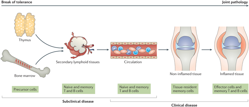
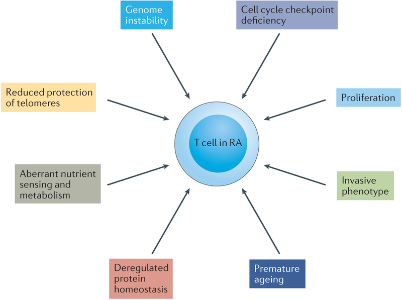
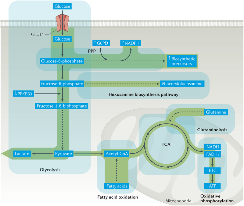
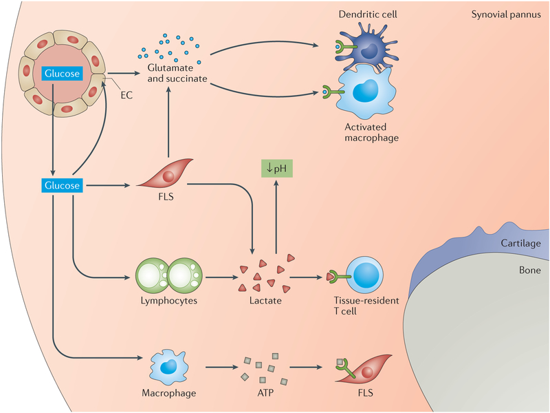
One of the fundamental traits of immune cells in rheumatoid arthritis (RA) is their ability to proliferate, a property shared with the joint-resident cells that form the synovial pannus. The building of biomass imposes high demands for energy and biosynthetic precursors, implicating metabolic control as a basic disease mechanism. During preclinical RA, when autoreactive T cells expand and immunological tolerance is broken, the main sites of disease are the secondary lymphoid tissues. Naive CD4+ T cells from patients with RA have a distinct metabolic signature, characterized by dampened glycolysis, low ATP levels and enhanced shunting of glucose into the pentose phosphate pathway. Equipped with high levels of NADPH and depleted of intracellular reactive oxygen species, such T cells hyperproliferate and acquire proinflammatory effector functions. During clinical RA, immune cells coexist with stromal cells in the acidic milieu of the inflamed joint. This microenvironment is rich in metabolic intermediates that are released into the extracellular space to shape cell-cell communication and the functional activity of tissue-resident cells. Increasing awareness of how metabolites regulate signalling pathways, guide post-translational modifications and condition the tissue microenvironment will help to connect environmental factors with the pathogenic behaviour of T cells in RA.


Conflict of interest statement

Competing interests statement

The authors declare no competing interests.

Figures

Figure 1
Figure 1. The disease process of rheumatoid arthritis in different tissue environments.
The disease process of rheumatoid arthritis (RA) involves multiple tissue environments and extends over several decades. An early event is the breakdown of immunologic tolerance, which occurs in lymphoid tissues. After expansion and maturation of autoreactive lymphocytes, autoantigens are encountered in peripheral tissues, eventually leading to the formation of tertiary lymphoid microstructures and chronic destructive inflammation. The inflamed synovial membrane and the disrupted tissue repair response represent the end stage of RA.
Figure 2
Figure 2. Emerging hallmarks of T cells in rheumatoid arthritis.
Core hallmarks of T cells in rheumatoid arthritis (RA) are their ability to massively proliferate and to differentiate into proinflammatory effector cells. Several changes in the basic biologic pathways listed here distinguish T cells in healthy individuals from those in patients with RA and enable such T cells to deviate from their protective role to an autoinflammatory one. The molecular defects underlying pathogenic T-cell behaviour are currently being discovered; among them is the reprogramming of cellular metabolism, which fuels the functional capabilities of arthritogenic T cells.
Figure 3
Figure 3. Glucose shunting into the pentose phosphate pathway in T cells in RA.
Glycolytic breakdown of glucose in T cells in rheumatoid arthritis (RA) is reduced as a result of diminished activity of the regulatory enzyme 6-phosphofructo-2-kinase/fructose-2,6-bisphophastase 3 (PFKFB3), which curbs ATP production. With increased activity of glucose-6-phosphate dehydrogenase (G6PD), glucose is shunted to the pentose phosphate pathway (PPP), yielding high amounts of the electron donor NADPH and generation of biosynthetic precursors. Cellular stores of glutathione are shifted towards the reduced form, cellular levels of reactive oxygen species (ROS) are depleted and cellular oxidant signalling is impaired. ETC, electron transport chain; GLUT1, glucose transporter type 1; TCA, tricarboxylic acid cycle.
Figure 4
Figure 4. Metabolic intermediates in the RA joint.
The synovial pannus in the joints of patients with rheumatoid arthritis (RA) is a hypermetabolic lesion that demands high amounts of nutrients and oxygen to fulfil the energy and biosynthetic needs of its proliferative cells. The presence of glucose enables rapid and adaptive production of ATP, even under hypoxic conditions. Metabolic products such as lactate and ATP are released into the extracellular space, where they promote cell–cell communication and regulatory control. Lactate acidifies the tissue microenvironment and might directly contribute to cellular injury. With high levels of mitochondrial activity in tissue-resident and invasive cells, intermediates of the tricarboxylic acid cycle such as succinate and glutamate are secreted into the extracellular space. Signals transduced through specialized receptors (such as hydroxycarboxylic acid receptor 1 for lactate and succinate receptor 1 for succinate) regulate the functions of cells that sense extracellular metabolites. Here, metabolites serve as signalling molecules in cell–cell communication and in microenvironmental surveillance.

Similar articles

Cited by

References

    1. Rantapaa-Dahlqvist S et al. Antibodies against cyclic citrullinated peptide and IgA rheumatoid factor predict the development of rheumatoid arthritis. Arthritis Rheum. 48, 2741–2749 (2003). - PubMed
    1. Majka DS & Holers VM Can we accurately predict the development of rheumatoid arthritis in the preclinical phase? Arthritis Rheum. 48, 2701–2705 (2003). - PubMed
    1. Arbuckle MR et al. Development of autoantibodies before the clinical onset of systemic lupus erythematosus. N Engl J Med. 349, 1526–1533 (2003). - PubMed
    1. Kimpimaki T & Knip M Disease-associated autoantibodies as predictive markers of type 1 diabetes mellitus in siblings of affected children. J Pediatr Endocrinol Metab. 14 Suppl 1, 575–587 (2001). - PubMed
    1. Knip M et al. Prediction of type 1 diabetes in the general population. Diabetes Care. 33, 1206–1212 (2010). - PMC - PubMed

Substances