

Novel insights into the interaction of UBA5 with UFM1 via a UFM1-interacting sequence
- PMID: 28360427
- PMCID: PMC5428781
- DOI: 10.1038/s41598-017-00610-0
Novel insights into the interaction of UBA5 with UFM1 via a UFM1-interacting sequence
Abstract
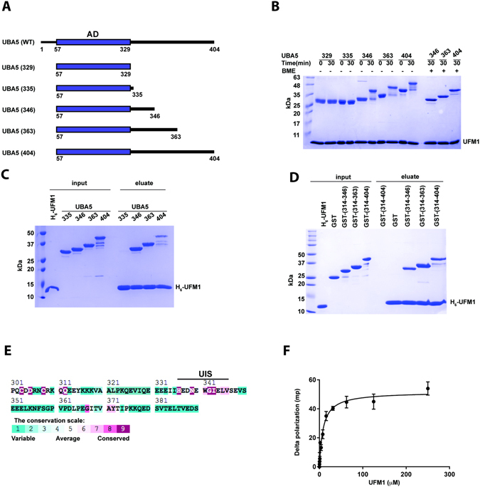
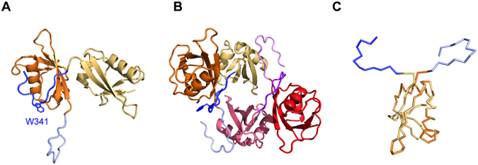
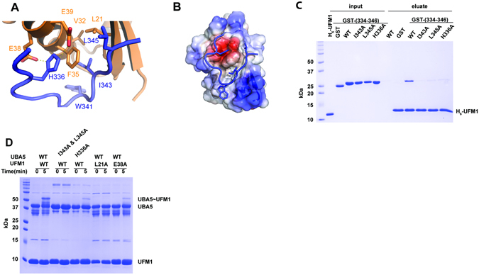
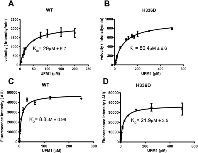
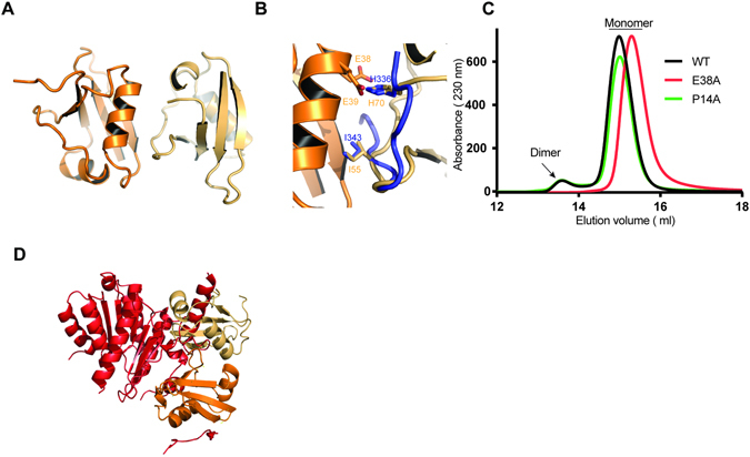
The modification of proteins by ubiquitin-fold modifier 1 (UFM1) is implicated in many human diseases. Prior to conjugation, UFM1 undergoes activation by its cognate activating enzyme, UBA5. UBA5 is a non-canonical E1 activating enzyme that possesses an adenylation domain but lacks a distinct cysteine domain. Binding of UBA5 to UFM1 is mediated via an amino acid sequence, known as the UFM1-interacting sequence (UIS), located outside the adenylation domain that is required for UFM1 activation. However, the precise boundaries of the UIS are yet not clear and are still under debate. Here we revisit the interaction of UFM1 with UBA5 by determining the crystal structure of UFM1 fused to 13 amino acids of human UBA5. Using binding and activity assays, we found that His 336 of UBA5, previously not reported to be part of the UIS, occupies a negatively charged pocket on UFM1's surface. This His is involved in UFM1 binding and if mutated perturbs activation of UFM1. Surprisingly, we also found that the interaction between two UFM1 molecules mimics how the UIS binds UFM1. Specifically, UFM1 His 70 resembles UBA5 His336 and enters a negatively charged pocked on the other UFM1 molecule. Our results refine our understanding of UFM1-UBA5 binding.
Conflict of interest statement
The authors declare that they have no competing interests.
Figures
Similar articles
-
Structural and Functional Analysis of a Novel Interaction Motif within UFM1-activating Enzyme 5 (UBA5) Required for Binding to Ubiquitin-like Proteins and Ufmylation.J Biol Chem. 2016 Apr 22;291(17):9025-41. doi: 10.1074/jbc.M116.715474. Epub 2016 Feb 29. J Biol Chem. 2016. PMID: 26929408 Free PMC article.
-
An N-Terminal Extension to UBA5 Adenylation Domain Boosts UFM1 Activation: Isoform-Specific Differences in Ubiquitin-like Protein Activation.J Mol Biol. 2019 Feb 1;431(3):463-478. doi: 10.1016/j.jmb.2018.10.007. Epub 2018 Nov 6. J Mol Biol. 2019. PMID: 30412706
-
Trans-binding of UFM1 to UBA5 stimulates UBA5 homodimerization and ATP binding.FASEB J. 2018 May;32(5):2794-2802. doi: 10.1096/fj.201701057R. Epub 2018 Jan 8. FASEB J. 2018. PMID: 29295865
-
Decrypting UFMylation: How Proteins Are Modified with UFM1.Biomolecules. 2020 Oct 14;10(10):1442. doi: 10.3390/biom10101442. Biomolecules. 2020. PMID: 33066455 Free PMC article. Review.
-
UFMylation: A Unique & Fashionable Modification for Life.Genomics Proteomics Bioinformatics. 2016 Jun;14(3):140-146. doi: 10.1016/j.gpb.2016.04.001. Epub 2016 May 20. Genomics Proteomics Bioinformatics. 2016. PMID: 27212118 Free PMC article. Review.
Cited by
-
UFM1 E3 ligase promotes recycling of 60S ribosomal subunits from the ER.Nature. 2024 Mar;627(8003):445-452. doi: 10.1038/s41586-024-07073-0. Epub 2024 Feb 21. Nature. 2024. PMID: 38383785 Free PMC article.
-
Structural basis for UFM1 transfer from UBA5 to UFC1.Nat Commun. 2021 Sep 29;12(1):5708. doi: 10.1038/s41467-021-25994-6. Nat Commun. 2021. PMID: 34588452 Free PMC article.
-
Allelic strengths of encephalopathy-associated UBA5 variants correlate between in vivo and in vitro assays.Elife. 2023 Dec 11;12:RP89891. doi: 10.7554/eLife.89891. Elife. 2023. PMID: 38079206 Free PMC article.
-
The UFMylation System in Proteostasis and Beyond.Trends Cell Biol. 2019 Dec;29(12):974-986. doi: 10.1016/j.tcb.2019.09.005. Epub 2019 Nov 6. Trends Cell Biol. 2019. PMID: 31703843 Free PMC article. Review.
-
UFM1-Activating Enzyme 5 (Uba5) Requires an Extension to Get the Job Done Right.J Mol Biol. 2019 Feb 1;431(3):479-482. doi: 10.1016/j.jmb.2018.11.017. Epub 2018 Nov 17. J Mol Biol. 2019. PMID: 30458173 Free PMC article. No abstract available.
References
Publication types
MeSH terms
Substances
LinkOut - more resources
Full Text Sources
Other Literature Sources
Molecular Biology Databases
