

Pulmonary mucormycosis diagnosed by convex probe endobronchial ultrasound-guided fine needle aspiration of cavity wall
- PMID: 28360470
- PMCID: PMC5351364
- DOI: 10.4103/0970-2113.201320
Pulmonary mucormycosis diagnosed by convex probe endobronchial ultrasound-guided fine needle aspiration of cavity wall
Abstract
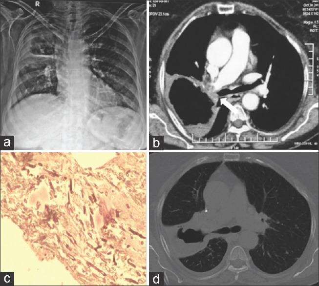
Pulmonary mucormycosis is an opportunistic fungal infection in immunocompromised individuals. It is difficult to diagnose as it requires tissue biopsy, and generally these patients are unfit to undergo invasive lung biopsies. We describe a novel technique in a case with uncontrolled diabetes mellitus with nonresolving pulmonary cavitary disease where convex probe endobronchial ultrasound (EBUS)-guided aspiration of lung cavity wall showed classical histopathological picture establishing the diagnosis of mucorale infection. EBUS being real-time, minimally invasive technique with minimal risk of complications, led to early diagnosis, and prompt treatment. This appears to be a novel diagnostic modality in pulmonary mucormycosis with minimal complications as compared with other biopsy methods with very high complication risk.
Keywords: Endobronchial ultrasound-guided fine needle aspiration; mucormycosis; pulmonary cavity.
Conflict of interest statement
There are no conflicts of interest.
Figures
Similar articles
-
Role of radial endobronchial ultrasound-guided transbronchial needle aspiration in the diagnosis of pulmonary nodules: Case report and literature review.Lung India. 2017 Jan-Feb;34(1):61-64. doi: 10.4103/0970-2113.197094. Lung India. 2017. PMID: 28144062 Free PMC article.
-
Complications of Convex-Probe Endobronchial Ultrasound-Guided Transbronchial Needle Aspiration: A Multi-Center Retrospective Study.Respir Care. 2016 Feb;61(2):243-8. doi: 10.4187/respcare.03838. Epub 2015 Nov 10. Respir Care. 2016. PMID: 26556895
-
Unusual diagnoses made by convex-probe endobronchial ultrasound-guided transbronchial needle aspiration.Pulmonology. 2018 Sep-Oct;24(5):300-306. doi: 10.1016/j.pulmoe.2017.12.004. Epub 2018 Apr 4. Pulmonology. 2018. PMID: 29627400 Review.
-
[Endobronchial ultrasound guided transbronchial needle aspiration (EBUS-TBNA) for the evaluation of the mediastinum].Kyobu Geka. 2007 Jul;60(8 Suppl):711-7. Kyobu Geka. 2007. PMID: 17763674 Japanese.
-
Scientific evidence and principles for the use of endobronchial ultrasound and transbronchial needle aspiration.Expert Rev Med Devices. 2011 Jul;8(4):493-513. doi: 10.1586/erd.11.14. Expert Rev Med Devices. 2011. PMID: 21728734 Review.
Cited by
-
The evolution of endobronchial ultrasound usage in modern era.Tuberk Toraks. 2023 Sep;71(3):299-307. doi: 10.5578/tt.20239711. Tuberk Toraks. 2023. PMID: 37740633 Free PMC article. Review.
-
Dental and Oral Manifestations of COVID-19 Related Mucormycosis: Diagnoses, Management Strategies and Outcomes.J Fungi (Basel). 2021 Dec 31;8(1):44. doi: 10.3390/jof8010044. J Fungi (Basel). 2021. PMID: 35049983 Free PMC article. Review.
-
Mucormycosis: Cytomorphological Spectrum in Fine-Needle Aspiration Cytology.J Cytol. 2024 Jan-Mar;41(1):47-52. doi: 10.4103/joc.joc_107_23. Epub 2023 Dec 28. J Cytol. 2024. PMID: 38282815 Free PMC article.
-
Challenges in the diagnosis and treatment of mucormycosis.Med Mycol. 2018 Apr 1;56(suppl_1):93-101. doi: 10.1093/mmy/myx101. Med Mycol. 2018. PMID: 29538730 Free PMC article. Review.
-
Role of Convex Probe Endobronchial Ultrasound in the Diagnosis and Treatment of Nonmalignant Diseases.Pulm Med. 2019 Jun 17;2019:6838439. doi: 10.1155/2019/6838439. eCollection 2019. Pulm Med. 2019. PMID: 31316830 Free PMC article. Review.
References
-
- Petrikkos G, Skiada A, Lortholary O, Roilides E, Walsh TJ, Kontoyiannis DP. Epidemiology and clinical manifestations of mucormycosis. Clin Infect Dis. 2012;54(Suppl 1):S23–34. - PubMed
-
- Glazer M, Nusair S, Breuer R, Lafair J, Sherman Y, Berkman N. The role of BAL in the diagnosis of pulmonary mucormycosis. Chest. 2000;117:279–82. - PubMed
-
- Hamilos G, Samonis G, Kontoyiannis DP. Pulmonary mucormycosis. Semin Respir Crit Care Med. 2011;32:693–702. - PubMed
LinkOut - more resources
Full Text Sources
Other Literature Sources
