Review
. 2017 Mar 16:8:32.
doi: 10.3389/fgene.2017.00032. eCollection 2017.

Jmjd6, a JmjC Dioxygenase with Many Interaction Partners and Pleiotropic Functions

Affiliations
Review

Jmjd6, a JmjC Dioxygenase with Many Interaction Partners and Pleiotropic Functions

Janice Kwok et al. Front Genet. .

Abstract

Lysyl hydroxylation and arginyl demethylation are post-translational events that are important for many cellular processes. The jumonji domain containing protein 6 (JMJD6) has been reported to catalyze both lysyl hydroxylation and arginyl demethylation on diverse protein substrates. It also interacts directly with RNA. This review summarizes knowledge of JMJD6 functions that have emerged in the last 15 years and considers how a single Jumonji C (JmjC) domain-containing enzyme can target so many different substrates. New links and synergies between the three main proposed functions of Jmjd6 in histone demethylation, promoter proximal pause release of polymerase II and RNA splicing are discussed. The physiological context of the described molecular functions is considered and recently described novel roles for JMJD6 in cancer and immune biology are reviewed. The increased knowledge of JMJD6 functions has wider implications for our general understanding of the JmjC protein family of which JMJD6 is a member.

Keywords: JmjC domain; PSR; chromatin; demethylation; hydroxylation; pausing; splicing; transcription.


Figures

FIGURE 1
FIGURE 1
Protein domains and motifs of Jmjd6. (A) Schematic presentation of the full-length JMJD6 protein (403 amino acids, UniProt ID Q6NYC1) with central JmjC domain (green), nuclear localization sites (NLS, blue), nuclear export signal (NES, red), predicted SUMOylation site (light blue), and C-terminal poly serine (pink). Fe2+ complexing residues are shown as purple balls (His187, Asp189, His273). Scale indicates the length of the 403 amino acid (aa) comprising protein (B) Sequence of an extended AT-hook-like motif in JMJD6. Shown on the top line is the consensus sequence of an extended AT hook (eAT) after (Filarsky et al., 2015). GRP indicates the glutamine-arginine-proline tripeptide core motif which is surrounded in close proximity by at least three basic leucine (L) and/or arginine (R) residues. X indicates any amino acid in a distance of 6–20 amino acids from the GRP core motif. Bottom line: peptide eAT-hook-like sequence of JMJD6 (residues 283–326). GRP core motif is depicted in blue, basic K and R amino acids in red. The central/canonical AT-hook sequence is underlined. Note that the JMJD6 eAT-hook sequence with basic amino acids (K/R) is only extended distally to the GRP core motif sequence. Thus, Jmjd6 appears to have a hybrid AT hook sequence motif as discussed in the text (see The Jmjd6 Protein – Basic Structural Features and Potential Functions).
FIGURE 2
FIGURE 2
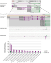
FANTOM5 analysis of the human locus encompassing the JMJD6 and METTL23 loci. FANTOM5 CAGE sequence data as depicted in the Zenbu browser and described by the (FANTOM Consortium and the RIKEN PMI and CLST (DGT) et al., 2014). Top line: shown are the Entrez hg 19 JMJD6 (purple arrow) and METTL23 (green arrow) transcriptional orientations. Sequence positions on human chromosome 17 are indicated on the top. Gencode v19 transcripts for JMJD6 (purple) and METTL23 (green) are shown below. Rectangles indicate exons and lines introns. Note: at the proximal and distal borders of the depicted JMJD6 and METTL23 loci the Zenbu browser screen view shows parts of the neighboring genes MXRA7 and SRSF2 in this gene dense genomic region. FANTOM5 CAGE and ENCODE CAGE sequence peaks are shown in the center. JMJD6 CAGE sequence peaks are shown in purple, METTL23 CAGE sequence peaks in green. Bottom graph: relative transcriptional start site (TSS/CAGE) expression data for JMJD6 (purple) and METLL23 (green) in blood cells from different donors. Relative TTS tag incidence refers to CAGE sequence tag counts across the locus which are quantified as normalized tags per million (TPM) as previously described (FANTOM Consortium and the RIKEN PMI and CLST (DGT) et al., 2014). CAGE sequence peaks for both genes are partially overlapping in eosinophils, neutrophils, and natural killer cells. The data shown are from the FANTOM5 database publically accessible at the Zenbu browser (http://fantom.gsc.riken.jp/5/) when searching for JMJD6 or METTL23.
FIGURE 3
FIGURE 3
Function of JMJD6 as a histone arginine demethylase. JMJD6 has been proposed to demethylate residues at histone H4 arginine 3 (H4R3) and histone H3 arginine 2 (H3R2). Arginine residues can be symmetrically (SDMA) or asymmetrically (ADMA) dimethylated. 2-oxoglutarate- (2OG) and oxygen (O2)-dependent demethylation of H4R3me2s and H3R2me2a by JMJD6 produces succinate, CO2, formaldehyde and monomethylated arginine residues. Both arginine histone marks, H4R3me2s and H3R2me2a are associated with transcriptionally inactive chromatin. Demethylation by JMJD6 to the monomethyl arginine (MMA) H4R3me1 or H3R2me1 histone marks is proposed to be associated with transcriptional activation.
FIGURE 4
FIGURE 4
JMJD6 as a regulator of RNA polymerase II promoter-proximal pause release. Mechanistic model after Liu et al. (2013) on how JMJD6 regulates transcriptional pause release through dual demethylation activity on H4R3me2s and the 7SK snRNA via interaction with BRD4 on anti-pause enhancers. Demethylation of the 5′-methyl cap of the 7SK snRNA by JMJD6 leads to the dissociation of the positive transcription elongation factor-b (P-TEFb) complex from the HEXIM1/2 polymerase II (Pol II) inhibitory complex. The P-TEFb complex (circled with a dashed green line) consists of the subunit proteins cyclin-dependent kinase 9 (CDK9) and cyclin T1/2 (CCNT1/2). Released P-TEFb phosphorylates serine 2 (Ser2-P) in the C-terminal heptapeptide repeat region of RNA-polymerase II (Pol II) and the negative elongation factor (NELF) which causes its dissociation from the DRB sensitivity-inducing factor (DSIF). Both phosphorylation events facilitate release of paused Pol II and efficient mRNA elongation. In the process of Pol II promoter-proximal pause release recruited JMD6 also demethylates H4R3me2s. Genomic DNA with nucleosome and anti-pause enhancer depicted as gray line; mRNA (yellow line); 7SK snRNA (blue line); BRD4, bromodomain-containing protein 4 (purple); DSIF, DRB sensitivity-inducing factor (blue); JMJD6, Jumonji domain containing protein 6 (red); Pol II, RNA-polymerase II (dark green); NELF, negative elongation factor (orange); P-TEFb subunits CDK9, cyclin-dependent kinase 9 (light green) and CCNT1/2, cyclin T1/2 (light green); HEXIM 1/2 dimer, hexamethylene bisacetamide inducible proteins 1 and 2 (violet); Ser2-P, phosphorylated serine 2 of Pol II and TSS, transcription start site; red lines indicating Jmjd6 mediated demethylation reactions.
FIGURE 5
FIGURE 5
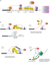
Functions of Jmjd6 in the regulation of splicing. (A) Possible role for JMJD6 in modulating binding of splicing regulatory proteins to splice recognition sites on pre-mRNAs. JMJD6 forms a trimeric complex with U2AF65/U2AF35 and has been demonstrated to modulate splicing by lysyl hydroxylation of U2AF65. JMJD6 interacts also with SR proteins that can bind to exon splicing enhancers (ESE) in proximity to 3′-splice acceptor sites. Examples of these SR proteins are LUC7L1, LUC7L2, LUC7L3 (CROP), SRSF11, and Acinus S’ (Heim et al., 2014). JMJD6 mediated hydroxylation of SR proteins might influence selection of exonic 3′-splice acceptor sites through stabilization or weakening of U2AF binding to polypyrimidine tracts. Shown are U1 snRNP binding to exonic 5′-splice donor sites, binding of splicing factor 1 (SF1) and SR protein complexes to intronic branch point sequences (BPS), and assembly of the U2 spliceosome components U2AF65 and U2AF35 together with JMJD6 and SR proteins at the polypyrimidine tract (Py-tract) and 3′-splice acceptor site. Examples of SR proteins that interact with JMJD6 are depicted in the box on the left. (B) Hypotheses on mechanisms how JMJD6 could regulate mRNA splicing (see Functions of Jmjd6 in RNA Processing – Regulation of Splicing). Hypothesis (1): Through JMJD6 mediated hydroxylation of SR proteins that bind to exonic splicing enhancers (ESE) exon skipping may be induced. In this case, SR protein hydroxylation by JMJD6 at a local 3′-splice acceptor site might suppress assembly of U2 snRNP components while preferential assembly of unmodified U2 splicing factors and SR proteins might occur at a 3′-splice acceptor site further downstream on the pre-mRNA. Hypothesis (2): Hydroxylation of U2 snRNP components and SR proteins by JMJD6 may induce lopping of pre-mRNAs into different secondary or tertiary RNA structures. JMJD6 mediated hydroxylation of SR proteins bound to exonic splicing silencers (ESS) may facilitate formation of pre-mRNA structures that inhibit splicing of upstream exons. The pre-mRNA folding platform cannot assemble and neighboring exons are not brought into proximity to facilitate splicing. Hypothesis (3): In this case JMJD6 mediated hydroxylation of U2 snRNP and SR proteins triggers the assembly of a platform at exonic splicing enhancers (ESE) that facilitates pre-mRNA looping and recruitment of co-factors needed for exon inclusion into spliced mRNAs. Hypothesis (4): Involvement of JMJD6 in co-transcriptional regulation of mRNA elongation and splicing. Hydroxylation of SR proteins by JMJD6 could regulate polymerase II (Pol II) transcriptional pause release and shuttling of SR proteins from the transcription start complex into spliceosomal complexes where these then regulate splicing reactions.
FIGURE 6
FIGURE 6
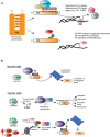
Roles of Jmjd6 in cancer. (A) JMJD6 regulates P53 functions by hydroxylation. Inactive P53 is normally locked in a closed confirmation which prevents its binding to target genes on genomic DNA. Acetylation by different acetyltransferases such as p300/CBP (blue) and the p300-CBP associated factor (PCAF, yellow) at C-terminal located lysines (K370-K382) affects P53 transcriptional activity by opening up its closed confirmation exposing its central DNA binding domain. Binding of P53 to gene promoters and recruitment of co-factors induces expression of target genes such as P21 and PUMA. Lysyl hydroxylation of P53K382 by JMJD6 has been described to suppress expression of P21 and PUMA by preventing P53 acetylation at this amino acid (Wang et al., 2014). (B) Hypothesis of how amplification of Jmjd6 in mouse mammary tumor models inhibits c-Myc induced apoptosis and enhances tumorigenesis. In normal cells (top row) where c-Myc (green) is not mutated, the E3 ubiquitin-protein ligase Mdm2 (Mouse double minute 2 homolog, purple) ubiquitinates p53 thereby leading to its proteosomal degradation. In cancer cells (middle row), upregulated expression of mutated c-Myc induces Cdkna2 (cyclin-dependent kinase inhibitor 2A, also known as p19/14ARF, blue) expression. Cdkna2 binds to Mdm2 and antagonizes its ubiquitin ligase function thereby leading to stabilization of p53. Upregulated p53 induces several important cellular processes linked to tumor suppression (indicated by arrows on the left) foremost induction of apoptosis. In advanced MMTV-Myc induced tumors, the Jmjd6 gene copy number is amplified leading to elevated levels of Jmjd6 protein (red) expression (bottom row). Through its histone arginine demethylation activity, Jmjd6 demethylates the activating histone mark H4R3me2a at the Cdkn2a promoter leading to diminished expression of this Mdm2 inhibitor. P53 is subjected to proteosomal degradation leading consequently to a loss of its tumor suppression function. According to the model of Aprelikova et al. (2016) amplified Jmjd6 has pro-oncogenic functions through interference with p53 functions and elevated JMJD6 expression in tumor tissue is therefore an unfavorable prognostic marker for cancer prognosis.

References

    1. Adelman K., Lis J. T. (2012). Promoter-proximal pausing of RNA polymerase II: emerging roles in metazoans. Nat. Rev. Genet. 13 720–731. 10.1038/nrg3293 - DOI - PMC - PubMed
    1. Allis C. D., Berger S. L., Cote J., Dent S., Jenuwien T., Kouzarides T., et al. (2007). New nomenclature for chromatin-modifying enzymes. Cell 131 633–636. 10.1016/j.cell.2007.10.039 - DOI - PubMed
    1. Aprelikova O., Chen K., El Touny L. H., Brignatz-Guittard C., Han J., Qiu T., et al. (2016). The epigenetic modifier JMJD6 is amplified in mammary tumors and cooperates with c-Myc to enhance cellular transformation, tumor progression, and metastasis. Clin. Epigenetics 8:38 10.1186/s13148-016-0205-6 - DOI - PMC - PubMed
    1. Arur S., Uche U. E., Rezaul K., Fong M., Scranton V., Cowan A. E., et al. (2003). Annexin I is an endogenous ligand that mediates apoptotic cell engulfment. Dev. Cell 4 587–598. 10.1016/S1534-5807(03)00090-X - DOI - PubMed
    1. Barman-Aksozen J., Beguin C., Dogar A. M., Schneider-Yin X., Minder E. I. (2013). Iron availability modulates aberrant splicing of ferrochelatase through the iron- and 2-oxoglutarate dependent dioxygenase Jmjd6 and U2AF(65.). Blood Cells Mol. Dis. 51 151–161. 10.1016/j.bcmd.2013.05.008 - DOI - PubMed