. 2017 Feb;10(1):63-74.
doi: 10.1007/s12195-016-0467-2. Epub 2016 Sep 12.

Multiaxial Polarity Determines Individual Cellular and Nuclear Chirality

Affiliations

Multiaxial Polarity Determines Individual Cellular and Nuclear Chirality

Michael J Raymond Jr et al. Cell Mol Bioeng. 2017 Feb.

Abstract

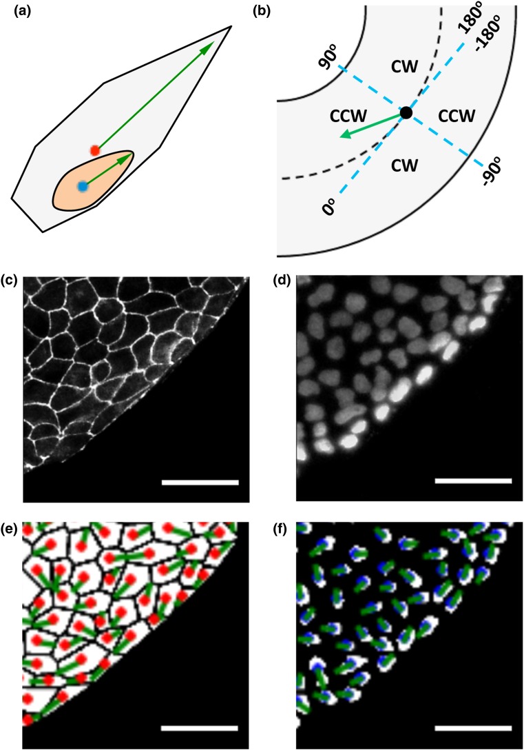
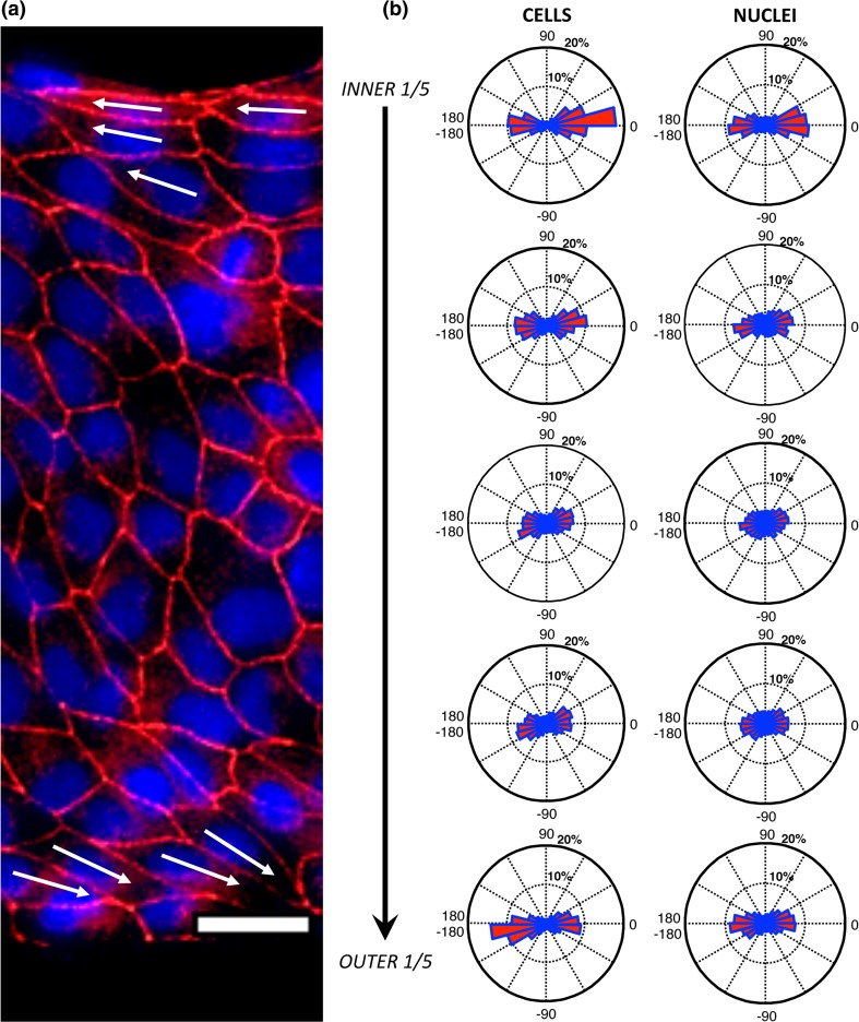
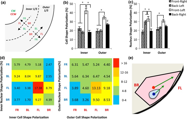
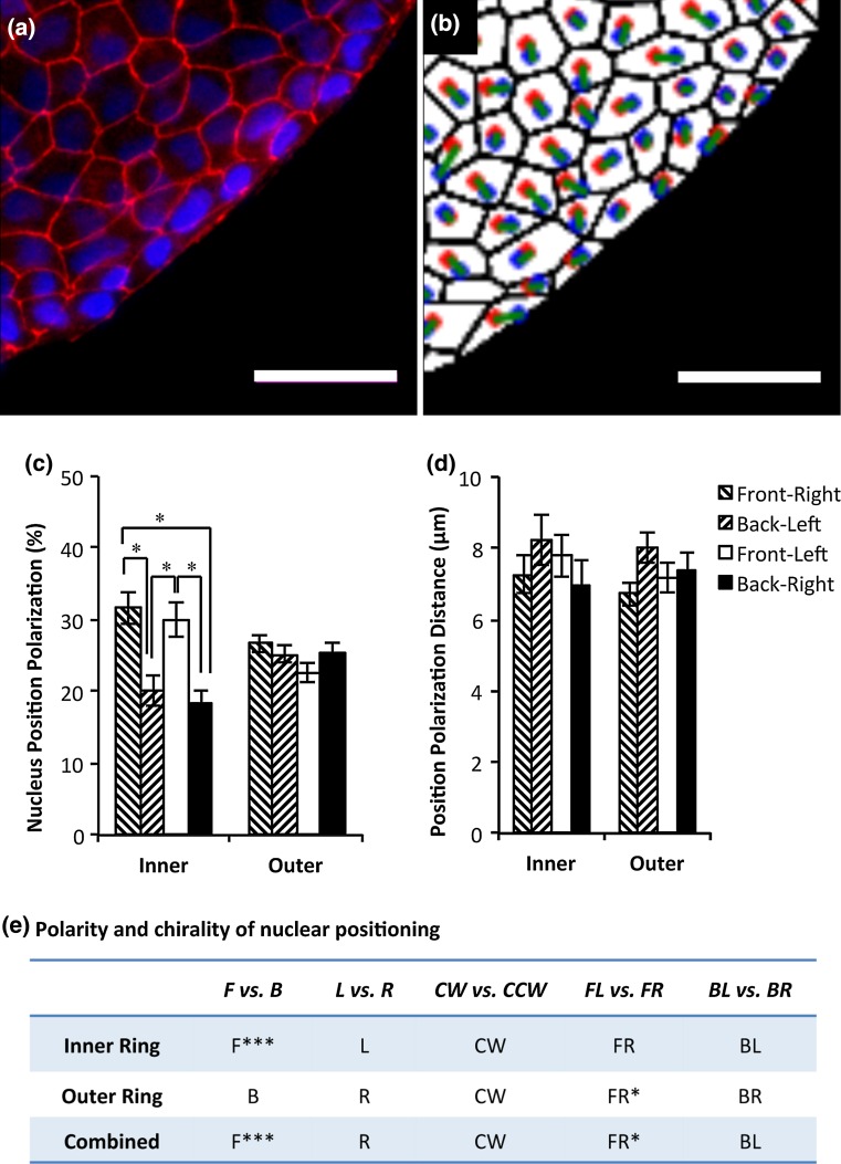
Intrinsic cell chirality has been implicated in the left-right (LR) asymmetry of embryonic development. Impaired cell chirality could lead to severe birth defects in laterality. Previously, we detected cell chirality with an in vitro micropatterning system. Here, we demonstrate for the first time that chirality can be quantified as the coordination of multiaxial polarization of individual cells and nuclei. Using an object labeling, connected component based method, we characterized cell chirality based on cell and nuclear shape polarization and nuclear positioning of each cell in multicellular patterns of epithelial cells. We found that the cells adopted a LR bias the boundaries by positioning the sharp end towards the leading edge and leaving the nucleus at the rear. This behavior is consistent with the directional migration observed previously on the boundary of micropatterns. Although the nucleus is chirally aligned, it is not strongly biased towards or away from the boundary. As the result of the rear positioning of nuclei, the nuclear positioning has an opposite chirality to that of cell alignment. Overall, our results have revealed deep insights of chiral morphogenesis as the coordination of multiaxial polarization at the cellular and subcellular levels.

Keywords: Cell Chirality; Cell Morphology; Cell Polarity; Nuclear Morphology.


Conflict of interest statement

CONFLICT OF INTEREST All authors, Michael J. Raymond, Poulomi Ray, Gurleen Kaur, Michael Fredericks, Ajay V. Singh, and Leo Q. Wan, declare that they have no conflict of interest.

Figures

Figure 1
Figure 1
Analysis of polarization biases and chirality in cell and nuclear shape. Scale = 50 μm. (a) The front of cells and nuclei is defined as the sharp end, as indicated by the green arrows (red dot for cell centroid and blue dot for nuclear centroid). (b) Definition of alignment angles in a polarization axis (blue dashed lines representing cylindrical coordinates). (c) A region of interest of ZO-1 (tight junctions) fluorescence image. (d) Nuclei with DAPI staining. (e) ZO-1 images are processed and cell shape polarization angles are calculated, with red dots for cell centroids and green lines for cell polarization direction. (f) Nuclear shape polarization was determined and shown with green lines. Blue dots represent nuclear centroids.
Figure 2
Figure 2
Variation of cell and nuclear shape polarization between the inner and outer boundary. Scale bar = 25 µm. (a) A region of a CCW ring stained for ZO-1 (red) and nuclei (blue). White arrows indicate cell shape polarization direction of representative cells at the boundaries. (b) Circular histograms show the distribution of shape polarization angles of cells (left) and nuclei (right) in 5 regions of the 38 rings that are equally divided in the radial distance from the inner to the outer ring.
Figure 3
Figure 3
Analysis of cell and nuclear shape polarization at boundaries. (a) Each boundary region adopts local coordinates with the cells facing the closest boundary, dividing the entire plane into four quadrants (FR: front-right, BL: back-left, FL: front-left, and BR: back-right). Among them, BR and FL (red) represent a CCW bias and FR and BL (green) represent a CW bias. (b) Analysis of cell shape polarization reveals a bias towards front left (FL) polarization at the inner ring, and FL and BR polarizations at the outer. * significantly different between quadrants, and # significantly different from the inner ring. (c) Nuclear shape polarization analysis reveals a strong bias towards FL and BL polarizations at boundaries. * significantly different between quadrants. (d) Heat maps between cell shape polarization and nuclear shape polarization at the inner (left) and outer (right) rings. (e) Cells tend to polarize themselves towards FL in regions near the micropatterned boundaries, while nuclei are biased to the left with no significant bias towards front or back (arrows: cellular or nuclear shape polarization, blue dot: nuclear centroid, and red dot: cell centroid).
Figure 4
Figure 4
Nuclear positioning polarization. Scale bars: 50 μm. (a) ZO-1 (red) and nuclei (blue) fluorescence images. (b) Nuclear positioning polarization, defined as a vector from cell centroid to nucleus centroid, was determined from image analysis. (red: cell centroids, blue: nucleus centroids, green: nucleus positioning vector connecting two centroids with a cell). (c) Analysis of nuclear positioning polarization angles. (d) Analysis of nuclear positioning distances. (e) Determining polarity and chirality of nuclear positioning inside the cells at the patterned boundaries. * Significantly different at p < 0.05 and ** Significantly different at p < 0.01.

Similar articles

Cited by

References

    1. Andrew DJ, Ewald AJ. Morphogenesis of epithelial tubes: insights into tube formation, elongation, and elaboration. Dev. Biol. 2010;341:34–55. doi: 10.1016/j.ydbio.2009.09.024. - DOI - PMC - PubMed
    1. Aylsworth AS. Clinical aspects of defects in the determination of laterality. Am. J. Med. Genet. 2001;101:345–355. doi: 10.1002/ajmg.1219. - DOI - PubMed
    1. Balcarova-Stander J, Pfeiffer SE, Fuller SD, Simons K. Development of cell surface polarity in the epithelial Madin–Darby canine kidney (MDCK) cell line. EMBO J. 1984;3:2687–2694. - PMC - PubMed
    1. Callander DC, Alcorn MR, Birsoy B, Rothman JH. Natural reversal of left-right gut/gonad asymmetry in C. elegans males is independent of embryonic chirality. Genesis. 2014;52:581–587. doi: 10.1002/dvg.22762. - DOI - PMC - PubMed
    1. Chen TH, Hsu JJ, Zhao X, Guo C, Wong MN, Huang Y, Li Z, Garfinkel A, Ho CM, Tintut Y, Demer LL. Left-right symmetry breaking in tissue morphogenesis via cytoskeletal mechanics. Circ. Res. 2012;110:551–559. doi: 10.1161/CIRCRESAHA.111.255927. - DOI - PMC - PubMed

LinkOut - more resources