

Role of dynamic computed tomography scans in patients with congenital craniovertebral junction malformations
- PMID: 28361020
- PMCID: PMC5359763
- DOI: 10.5312/wjo.v8.i3.271
Role of dynamic computed tomography scans in patients with congenital craniovertebral junction malformations
Abstract
Aim: To evaluate the role of dynamic computed tomography (CT) scan imaging in diagnosing craniovertebral junction (CVJ) instability in patients with congenital CVJ malformations.
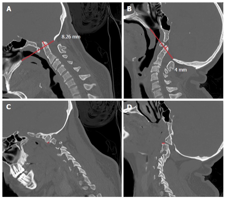
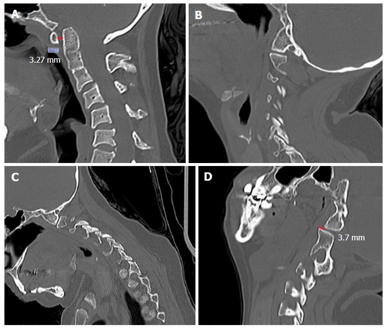
Methods: Patients with symptomatic congenital CVJ malformations who underwent posterior fossa decompression and had a preoperative dynamic CT scan in flexion and extended position were included in this study. Measurements of the following craniometrical parameters were taken in flexed and extended neck position: Atlanto-dental interval (ADI), distance of the odontoid tip to the Chamberlain's line, and the clivus-canal angle (CCA). Assessment of the facet joints congruence was also performed in both positions. Comparison of the values obtained in flexion and extension were compared using a paired Student's t-test.
Results: A total of ten patients with a mean age of 37.9 years were included. In flexion imaging, the mean ADI was 1.76 mm, the mean CCA was 125.4° and the mean distance of the odontoid tip to the Chamberlain's line was + 9.62 mm. In extension, the mean ADI was 1.46 mm (P = 0.29), the mean CCA was 142.2° (P < 0.01) and the mean distance of the odontoid tip to the Chamberlain's line was + 7.11 mm (P < 0.05). Four patients (40%) had facetary subluxation demonstrated in dynamic imaging, two of them with mobile subluxation (both underwent CVJ fixation). The other two patients with a fixed subluxation were not initially fixed. One patient with atlantoaxial assimilation and C23 fusion without initial facet subluxation developed a latter CVJ instability diagnosed with a dynamic CT scan. Patients with basilar invagination had a lower CCA variation compared to the whole group.
Conclusion: Craniometrical parameters, as well as the visualization of the facets location, may change significantly according to the neck position. Dynamic imaging can provide additional useful information to the diagnosis of CVJ instability. Future studies addressing the relationship between craniometrical changes and neck position are necessary.
Keywords: Basilar invagination; Chiari malformation; Craniovertebral junction; Dynamic imaging; Treatment.
Conflict of interest statement
Conflict-of-interest statement: The authors declare no conflicts of interest regarding this manuscript.
Figures

References
-
- Joaquim AF. Basilar invagination. J Neurosurg Pediatr. 2012;10:355; author reply 355–356. - PubMed
-
- Shah A, Goel A. Clival dysgenesis associated with Chiari Type 1 malformation and syringomyelia. J Clin Neurosci. 2010;17:400–401. - PubMed
-
- Goel A. Basilar invagination, Chiari malformation, syringomyelia: a review. Neurol India. 2009;57:235–246. - PubMed
LinkOut - more resources
Full Text Sources
Other Literature Sources
