. 2017 Aug;369(2):273-286.
doi: 10.1007/s00441-017-2598-8. Epub 2017 Mar 30.

Macrophage type modulates osteogenic differentiation of adipose tissue MSCs

Affiliations

Macrophage type modulates osteogenic differentiation of adipose tissue MSCs

Yang Zhang et al. Cell Tissue Res. 2017 Aug.

Abstract

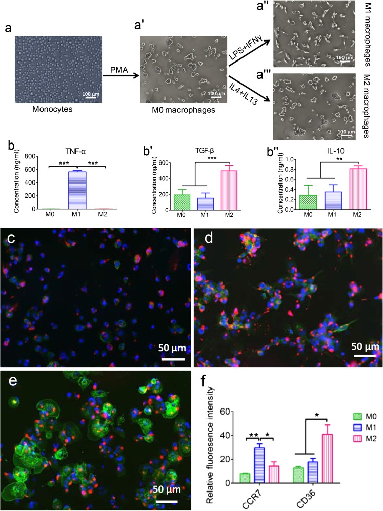
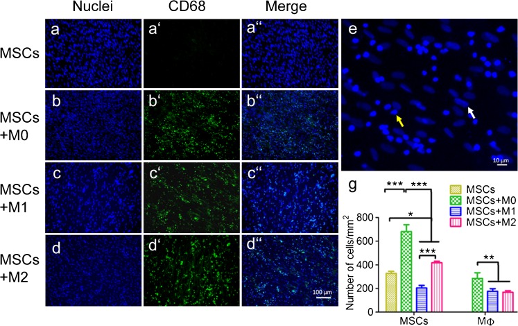
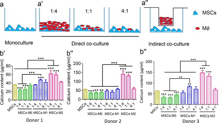
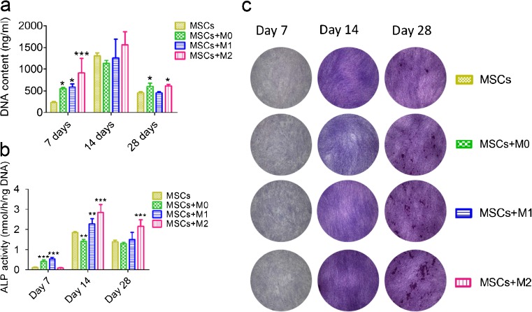
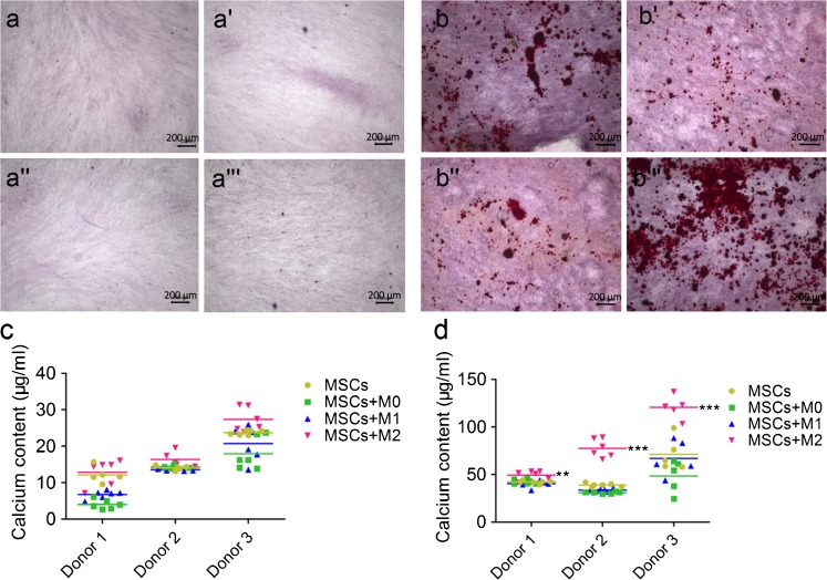
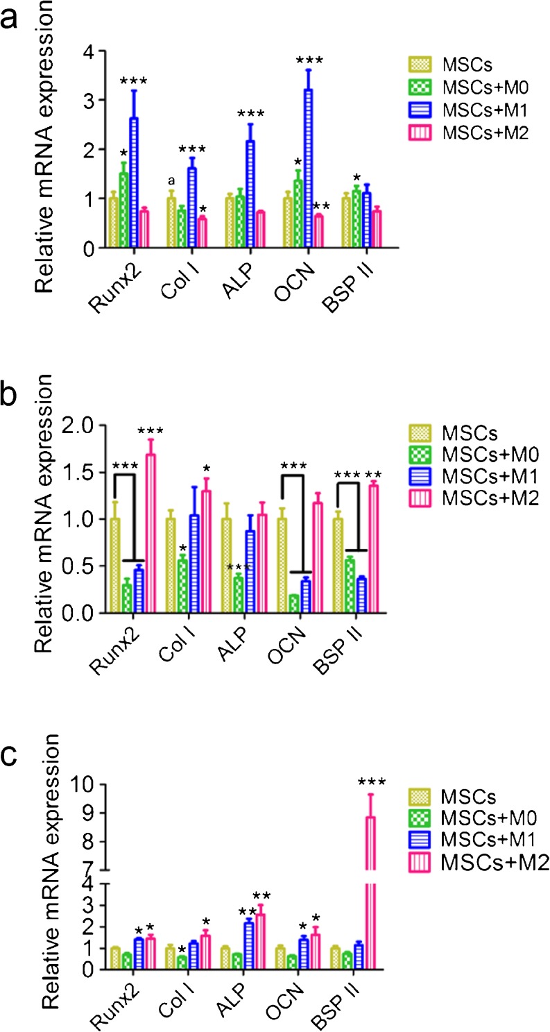
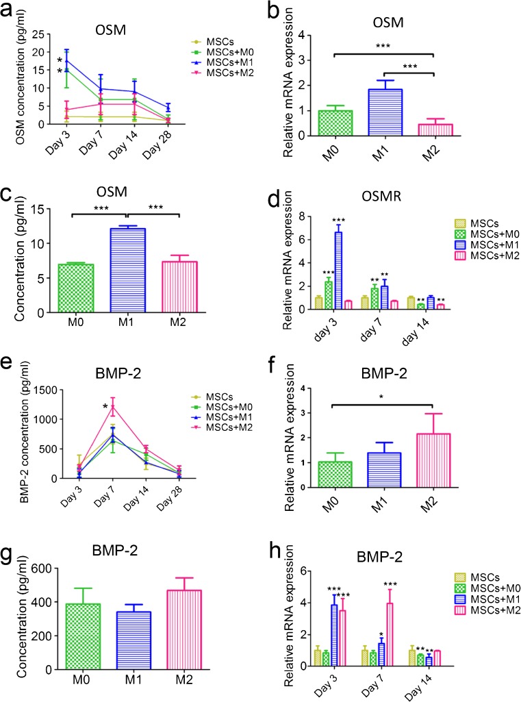
Since the reconstruction of large bone defects remains a challenge, knowledge about the biology of bone healing is desirable to develop novel strategies for improving the treatment of bone defects. In osteoimmunology, macrophages are the central component in the early stage of physiological response after bone injury and bone remodeling in the late stage. During this process, a switch of macrophage phenotype from pro-inflammatory (M1) to anti-inflammatory (M2) is observed. An appealing option for bone regeneration would be to exploit this regulatory role for the benefit of osteogenic differentiation of osteoprogenitor cells (e.g., mesenchymal stem cells; MSCs) and to eventually utilize this knowledge to improve the therapeutic outcome of bone regenerative treatment. In view of this, we focused on the in vitro interaction of different macrophage subtypes with adipose tissue MSCs to monitor the behavior (i.e. proliferation, differentiation and mineralization) of the latter in dedicated co-culture models. Our data show that co-culture of MSCs with M2 macrophages, but not with M1 macrophages or M0 macrophages, results in significantly increased MSC mineralization caused by soluble factors. Specifically, M2 macrophages promoted the proliferation and osteogenic differentiation of MSCs, while M0 and M1 macrophages solely stimulated the osteogenic differentiation of MSCs in the early and middle stages during co-culture. Secretion of the soluble factors oncostatin M (OSM) and bone morphogenetic protein 2 (BMP-2) by macrophages showed correlation with MSC gene expression levels for OSM-receptor and BMP-2, suggesting the involvement of both signaling pathways in the osteogenic differentiation of MSCs.

Keywords: Cell culture model; MSC; Macrophage; Osteogenic differentiation.


Conflict of interest statement

Conflict of interest

The authors have no conflict of interest to disclose.

Funding

This study was financially supported by The Netherlands Organization for Health Research and Development (ZonMw, project number 40-41400-98-1401) and China Scholarship Council (No. 2010622061).

Figures

Fig. 1
Fig. 1
The polarization method and cytokine expression of M0, M1 and M2 macrophages derived from THP-1 monocytes. The schematic figure shows the differentiation of monocytes into polarized macrophages and their morphological appearance (a a'''). TNF-α (b), TGF-β (b') and IL-10 (b'') production in M0, M1 and M2 macrophages conditioned medium were assessed by ELISA. M0 (c), M1 (d) and M2 (e) macrophages were stained with M1-marker CCR7 (red), M2-marker CD36 (green) and DAPI (blue), respectively. The relative fluorescence intensity of CCR7 and CD36 were quantified by ImageJ (f). Statistical analysis was performed by one-way ANOVA with Bonferroni’s test. *P ≤ 0.05; ** P ≤ 0.01, *** P ≤ 0.001
Fig. 2
Fig. 2
Immunostaining of different types of macrophages (MФ) co-cultured with MSCs. MSCs were monocultured or co-cultured with M0, M1 and M2 macrophages (1:4 ratio) for 4 weeks and stained with DAPI (blue, ad), pan-macrophage marker CD68 (green, a'd'), and then merged (a''d''). The number of macrophages and MSCs was counted based on shape of nuclei (e) and quantified (g). White arrow indicates the MSC while yellow arrow indicates the macrophage. Statistical analysis was performed by one-way ANOVA with Bonferroni’s test. n = 4, *P ≤ 0.05, ** P ≤ 0.01, *** P ≤ 0.001
Fig. 3
Fig. 3
Mineralization of MSCs directly co-cultured with M0, M1 and M2 macrophages. The schematic diagram shows the set-up of MSCs monoculture (a), direct co-culture (at different ratios; a') or indirect co-culture (at ratio of 1:4; a'') with M0, M1 and M2 macrophages respectively. Mineralization capacity of monoculture and direct co-culture from 3 donors was assessed by calcium content after 4 weeks in osteogenic medium (bb''). Statistical significance relative to controls (MSCs monoculture) and between groups was determined by two-way ANOVA with Bonferroni’s test correction, n = 5, **P ≤ 0.01, *** P ≤ 0.001. Asterisks on the top of the columns indicate significant differences from the MSCs control
Fig. 4
Fig. 4
Cell proliferation and ALP activity of MSCs indirectly co-cultured with M0, M1 and M2 macrophages. MSCs were monocultured and indirectly co-cultured with three types of macrophages and their proliferation was determined by DNA content assay (a) and their osteogenic differentiation was determined by ALP-activity assay (b) and ALP staining (c). Statistical analysis was performed by one-way ANOVA with Dunnett’s post-test. n = 4, * P ≤ 0.05, ** P ≤ 0.01, *** P ≤ 0.001
Fig. 5
Fig. 5
Mineralization of MSCs indirectly co-cultured with M0, M1 and M2 macrophages. MSCs monocultured (a, b) or indirectly co-cultured with M0 (a', b'), M1 (a', b'') and M2 (a''', b''') macrophages at a ratio of 1:4 were stained with alizarin red after 2 weeks (aa''') and 4 weeks (bb'''), respectively. Calcium content of MSCs (3 different donors) monocultured or indirectly co-cultured with three types of macrophages in the osteogenic medium was determined after 2 weeks (c) and 4 weeks (d), respectively. Statistical analysis was performed by one-way ANOVA with Dunnett’s post-test. n = 5, *P ≤ 0.05, ** P ≤ 0.01, * **P ≤ 0.001
Fig. 6
Fig. 6
Gene expression of osteogenic markers by MSCs indirectly co-cultured with M0, M1 and M2 macrophages. MSCs were indirectly co-cultured with three types of macrophages and their key osteogenic gene expression was determined by RT-PCR after 3 days (a), 7 days (b) and 14 days (c). Statistical analysis was performed by one-way ANOVA with Dunnett’s post-test. n = 4, *P ≤ 0.05, ** P ≤ 0.01, *** p ≤ 0.001
Fig. 7
Fig. 7
Secretion of key osteogenesis-related proteins in MSCs monoculture, MSCs and macrophages indirect co-cultures and macrophages conditioned medium. OSM secretion in macrophages and MSCs co-culture medium was assessed by ELISA (a). OSM gene expression of M0, M1 and M2 macrophages was determined by RT-PCR (b) and its protein secretion in conditioned medium was determined by ELISA (c). OSM receptor (OSMR) gene expression in MSCs was determined by RT-PCR (d). BMP-2 secretion in macrophages and MSCs co-culture medium was assessed by ELISA (e). BMP-2 gene expression of M0, M1 and M2 macrophages was determined by RT-PCR (f) and its protein secretion in conditioned medium was determined by ELISA (g). BMP-2 gene expression in MSCs was determined by RT-PCR (h). One-way ANOVA with Bonferroni’s correction was performed for comparison of OSM and BMP-2 gene expression and protein secretion between different types of macrophages. One-way ANOVA with Dunnett’s post-test correction was performed for comparison of OSM and BMP-2 concentration, OSMR and BMP-2 gene expression in MSCs. n = 4, *P ≤ 0.05, ** P ≤ 0.01, *** P ≤ 0.001

Similar articles

Cited by

References

    1. Alexander KA, Chang MK, Maylin ER, Kohler T, Müller R, Wu AC, Van Rooijen N, Sweet MJ, Hume DA, Raggatt LJ. Osteal macrophages promote in vivo intramembranous bone healing in a mouse tibial injury model. J Bone Miner Res. 2011;26(7):1517–1532. doi: 10.1002/jbmr.354. - DOI - PubMed
    1. Chen Z, Mao X, Tan L, Friis T, Wu C, Crawford R, Xiao Y. Osteoimmunomodulatory properties of magnesium scaffolds coated with β-tricalcium phosphate. Biomaterials. 2014;35(30):8553–8565. doi: 10.1016/j.biomaterials.2014.06.038. - DOI - PubMed
    1. Chen Z, Wu C, Gu W, Klein T, Crawford R, Xiao Y. Osteogenic differentiation of bone marrow MSCs by β-tricalcium phosphate stimulating macrophages via BMP2 signalling pathway. Biomaterials. 2014;35(5):1507–1518. doi: 10.1016/j.biomaterials.2013.11.014. - DOI - PubMed
    1. Cho SW, Soki FN, Koh AJ, Eber MR, Entezami P, Park SI, van Rooijen N, McCauley LK. Osteal macrophages support physiologic skeletal remodeling and anabolic actions of parathyroid hormone in bone. Proc Natl Acad Sci U S A. 2014;111(4):1545–1550. doi: 10.1073/pnas.1315153111. - DOI - PMC - PubMed
    1. Dimitriou R, Tsiridis E, Giannoudis PV. Current concepts of molecular aspects of bone healing. Injury. 2005;36(12):1392–1404. doi: 10.1016/j.injury.2005.07.019. - DOI - PubMed

Publication types