Review
. 2017 Mar;5(2):10.1128/microbiolspec.tbtb2-0027-2016.
doi: 10.1128/microbiolspec.TBTB2-0027-2016.

DNA Replication in Mycobacterium tuberculosis

Affiliations
Review

DNA Replication in Mycobacterium tuberculosis

Zanele Ditse et al. Microbiol Spectr. 2017 Mar.

Abstract

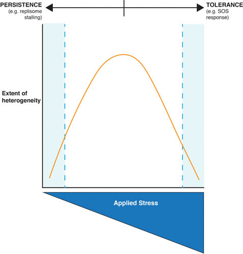
Faithful replication and maintenance of the genome are essential to the ability of any organism to survive and propagate. For an obligate pathogen such as Mycobacterium tuberculosis that has to complete successive cycles of transmission, infection, and disease in order to retain a foothold in the human population, this requires that genome replication and maintenance must be accomplished under the metabolic, immune, and antibiotic stresses encountered during passage through variable host environments. Comparative genomic analyses have established that chromosomal mutations enable M. tuberculosis to adapt to these stresses: the emergence of drug-resistant isolates provides direct evidence of this capacity, so too the well-documented genetic diversity among M. tuberculosis lineages across geographic loci, as well as the microvariation within individual patients that is increasingly observed as whole-genome sequencing methodologies are applied to clinical samples and tuberculosis (TB) disease models. However, the precise mutagenic mechanisms responsible for M. tuberculosis evolution and adaptation are poorly understood. Here, we summarize current knowledge of the machinery responsible for DNA replication in M. tuberculosis, and discuss the potential contribution of the expanded complement of mycobacterial DNA polymerases to mutagenesis. We also consider briefly the possible role of DNA replication-in particular, its regulation and coordination with cell division-in the ability of M. tuberculosis to withstand antibacterial stresses, including host immune effectors and antibiotics, through the generation at the population level of a tolerant state, or through the formation of a subpopulation of persister bacilli-both of which might be relevant to the emergence and fixation of genetic drug resistance.


Figures

FIGURE 1
FIGURE 1
A working model of the mycobacterial replisome. Schematic representation of the model replisome consisting of the PolIII core polymerase, the homodimeric β2-sliding clamp, the τ3δδ′ clamp-loader complex, DnaB helicase (red hexamer), DnaG primase (blue), PolI (pink) DNA ligase (purple), and SSB (orange). Recent biochemical evidence suggests that, in M. tuberculosis, the ε proofreader forms part of the core replicase together with the β2 and α subunits (42). As noted in the main text, the precise stoichiometry and architecture of the mycobacterial replisome remain to be established; similarly, it is not known whether the mycobacterial replisome functions as a di- or tripolymerase system, nor whether DnaE2 is able to access the replisome under non-DNA-damaging conditions in the absence of ImuB and ImuA′ accessory factors.
FIGURE 2
FIGURE 2
Subcomplex division in the bacterial replisome. The replisome contains three catalytic centers: core, clamp loader, and helicase-primase. The core complex and clamp-loader complex assemble into a larger, stable complex termed Pol III*. Together with the β clamp, they form the Pol III holoenzyme. The DnaB helicase and DnaG primase form a transient complex to synthesize primers on the lagging strand. Modified with permission from the Annual Review of Biochemistry, Volume 74 © 2005 by Annual Reviews, http://www.annualreviews.org
FIGURE 3
FIGURE 3
Structure of the C-family polymerases. (A) Computational model of M. tuberculosis DnaE1 based on the crystal structure of T. aquaticus PolIII. Different domains indicated in separate colors (C-terminal domains not shown). (B) Domain organization in the different polymerase families. The DnaE families are defined by the presence of the C-terminal domains, whereas PolC forms a distinct class where an ε-like exonuclease domain is inserted into the PHP domain.
FIGURE 4
FIGURE 4
Population heterogeneity as a function of the applied stress. The cartoon summarizes the notion that the degree (or strength) of applied stress might determine the extent of phenotypic heterogeneity within a specific (myco)bacterial population. So, as the applied stress (e.g., genotoxin, antibiotic, nutrient deprivation, pH, oxygen starvation) increases toward a critical point or concentration (which will differ for each stress), the degree of heterogeneity within the population increases. Beyond that critical point (the vertex of the parabola), the result is more likely to be manifest as a general, regulated response at the population level; this has the effect of reducing the extent of heterogeneity within the population. At each extreme (low/absent stress versus high/severe stress), the degree of heterogeneity approaches a minimum. Importantly, for conditions under which both the applied stress and the degree of heterogeneity are low, a small subpopulation of persister cells might enable survival, consistent with the framework proposed by Balaban and colleagues (177). At the other extreme—high/severe stress, low heterogeneity—any observed tolerance will exist at the population level, and will be mediated by a dominant regulatory mechanism(s), such as the LexA/RecA-dependent SOS response.

Similar articles

Cited by

References

    1. Ambur OH, Davidsen T, Frye SA, Balasingham SV, Lagesen K, Rognes T, Tønjum T. 2009. Genome dynamics in major bacterial pathogens. FEMS Microbiol Rev 33:453–470. 10.1111/j.1574-6976.2009.00173.x [PubMed] [PubMed] - DOI - PMC - PubMed
    1. Russell DG. 2016. The ins and outs of the Mycobacterium tuberculosis-containing vacuole. Cell Microbiol 18:1065–1069. 10.1111/cmi.12623 [PubMed] - DOI - PMC - PubMed
    1. Dartois V. 2014. The path of anti-tuberculosis drugs: from blood to lesions to mycobacterial cells. Nat Rev Microbiol 12:159–167. 10.1038/nrmicro3200 [PubMed] - DOI - PMC - PubMed
    1. Olive AJ, Sassetti CM. 2016. Metabolic crosstalk between host and pathogen: sensing, adapting and competing. Nat Rev Microbiol 14:221–234. 10.1038/nrmicro.2016.12 - DOI - PubMed
    1. Warner DF. 2014. Mycobacterium tuberculosis metabolism. Cold Spring Harb Perspect Med 5:5. [PubMed] - PMC - PubMed

Publication types

LinkOut - more resources