Review
. 2017 Aug;24(8):1390-1400.
doi: 10.1038/cdd.2017.47. Epub 2017 Mar 31.

Killers creating new life: caspases drive apoptosis-induced proliferation in tissue repair and disease

Affiliations
Review

Killers creating new life: caspases drive apoptosis-induced proliferation in tissue repair and disease

Caitlin E Fogarty et al. Cell Death Differ. 2017 Aug.

Abstract

Apoptosis is a carefully orchestrated and tightly controlled form of cell death, conserved across metazoans. As the executioners of apoptotic cell death, cysteine-dependent aspartate-directed proteases (caspases) are critical drivers of this cellular disassembly. Early studies of genetically programmed cell death demonstrated that the selective activation of caspases induces apoptosis and the precise elimination of excess cells, thereby sculpting structures and refining tissues. However, over the past decade there has been a fundamental shift in our understanding of the roles of caspases during cell death-a shift precipitated by the revelation that apoptotic cells actively engage with their surrounding environment throughout the death process, and caspases can trigger a myriad of signals, some of which drive concurrent cell proliferation regenerating damaged structures and building up lost tissues. This caspase-driven compensatory proliferation is referred to as apoptosis-induced proliferation (AiP). Diverse mechanisms of AiP have been found across species, ranging from planaria to mammals. In this review, we summarize the current knowledge of AiP and we highlight recent advances in the field including the involvement of reactive oxygen species and macrophage-like immune cells in one form of AiP, novel regulatory mechanisms affecting caspases during AiP, and emerging clinical data demonstrating the critical importance of AiP in cancer.


Conflict of interest statement

The authors declare no conflict of interest.

Figures
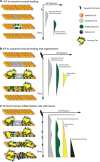
Figure 1
Figure 1
The Spectrum of AiP in wound healing, regeneration, and the development of cancer. On the left, shown are sheets of epithelial cells. On the right, the relative timing of caspase activity, AiP and the recruitment of immune cells as well as cancer cells (c) is illustrated. Based on the variety of caspase-dependent AiP mechanisms, it is clear that the phenomenon of AiP includes a spectrum of functions from simple transient wound healing, to more complex regeneration, to responding to chronic damage and inflammation. (a) Transient AiP is a self-limited proliferation in direct response to caspase activation in some apoptotic cells. (b) AiP associated with more significant or sustained wound healing often requires additional support including from the immune cells recruited by caspase-dependent signals such as extracellular ROS, but eventually resolves upon tissue repair. (c) Caspase-dependent AiP which occurs during ongoing or repeated damage, such as in chronic inflammatory diseases, can lead to an imbalance in cell death versus proliferation, leading to tissue dysplasia, hyperplasia, or possible neoplasia from the AiP stimulation of damaged cells containing new cancer-causing mutations
Figure 2
Figure 2
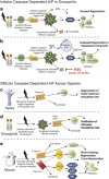
Mechanisms of caspase-dependent AiP. Apoptosis-induced proliferation may be induced by either initiator (a and b) or effector (ce) caspases. Activation of these caspases under certain conditions results in a variety of signaling cascades, producing a variety of growth factors and other mitogenic signals. (a and b) The Drosophila caspase Dronc is the only initiator caspase to date characterized as independently driving AiP. In transient regeneration (a), the active apoptosome including Dronc, triggers production of extracellular ROS and activates JNK, leading to production of the secreted mitogens Wingless (Wg) and Spitz (Spi). More sustained regeneration (b) requires additional input from recruited immune cells, signaling through the TNF homolog Eiger (Egr) and its receptor Grindelwald (Grnd), and possibly involves additional mitogens. (c–e) Effector caspases are implicated in AiP across the species, but examples shown here have confirmed the requirement of effector caspase activity. (c) In hydra, regeneration of an amputated head is dependent on caspase activity in the apoptotic cells at the regeneration site. These cells release the mitogen Wnt3. (d) The effector caspases Drice and DCP-1 are required for Hedgehog (Hh) production in Drosophila during AiP in maturing eye tissues. (e) In mice and human cell lines, following any number of injuries, including surgical resection, radiation or cytotoxic chemotherapies, caspase-3 cleaves a phospholipase A2 (iPLA2) triggering production of prostaglandin E2 (PGE2) which stimulates AiP. Caspase-3 also is required for VEGF-A production. Caspase-7 activates the kinase PKCδ, activating similar stress response pathways as seen in Drosophila
Figure 3
Figure 3
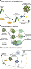
Regulation of caspases in AiP. Caspase activation may lead to apoptotic cell death or AiP. The exact mechanisms that direct caspase activity toward death or proliferative functions are not yet clear. When considering what is known regarding one of the better characterized pathways, the Dronc-dependent AiP in Drosophila epithelium, there are several possible regulatory mechanisms which could direct Dronc toward AiP. (a) Direct biochemical modification of caspases: Non-degradative mono-ubiquitinylation of Dronc inhibits both apoptotic and AiP activity. (b) Direction of caspases by adaptors or scaffolds: other non-apoptotic functions of Dronc rely on adaptors to localize the caspase to the non-apoptotic target. It is possible that Dronc-mediated AiP may be facilitated by an as of yet unidentified adaptor or scaffolding protein. (c) Feedback loops allow for amplification (green arrows) or inhibition (orange lines): positive feedback from JNK upregulating the pro-apoptotic factor hid increases Dronc activity and yields a robust AiP response. Negative feedback from EGFR signaling can inhibit Hid activity, potentially dampening the AiP response. As new downstream targets are uncovered for both initiator and effector caspase-dependent AiP, we will likely develop a better understanding of how a balance among these varying positive and inhibitory signals drive caspases towards a proliferative function
Figure 4
Figure 4
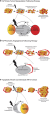
AiP in cancer and treatment failure. Emerging clinical studies have found that high levels of activated caspase-3 in tumors is associated with poor prognosis. Possible mechanisms by which caspase-mediated AiP could be contributing to tumorigenesis are modeled here. (a) Tumor repopulation following radiation or chemotherapy: cancer cells sensitive to the cytotoxic treatment initiate apoptosis and some cells trigger an AiP response through the production of PGE2 and other proliferative cytokines. Overall tumor bulk is initially reduced due to the apoptotic death, but the remaining cancer cells—including therapy-resistant cells—are stimulated via AiP to proliferate, repopulating the tumor. Over repeated cycles this could lead to extremely resistant tumors and eventual treatment failure. (b) Growth signals from dying tumor cells stimulate angiogenesis: caspase activation in dying tumor cells has been directly linked to VEGF-A production, a growth factor that promotes angiogenesis and neovascularization of the tumor. (c) Dying vascular endothelial cells also trigger AiP: caspase activation in the VE cells results in PGE2 production, which may stimulate further proliferation of the tumor cells

Similar articles

Cited by

References

    1. Haynie JL, Bryant PJ. The effects of X-rays on the proliferation dynamics of cells in the imaginal wing disc of Drosophila melanogaster. Dev Genes Evol 1977; 183: 85–100. - PubMed
    1. James AA, Bryant PJ. A quantitative study of cell death and mitotic inhibition in gamma-irradiated imaginal wing discs of Drosophila melanogaster. Radiat Res 1981; 87: 552–564. - PubMed
    1. Milan M, Campuzano S, Garcia-Bellido A. Developmental parameters of cell death in the wing disc of Drosophila. Proc Natl Acad Sci USA 1997; 94: 5691–5696. - PMC - PubMed
    1. Hwang JS, Kobayashi C, Agata K, Ikeo K, Gojobori T. Detection of apoptosis during planarian regeneration by the expression of apoptosis-related genes and TUNEL assay. Gene 2004; 333: 15–25. - PubMed
    1. Vlaskalin T, Wong CJ, Tsilfidis C. Growth and apoptosis during larval forelimb development and adult forelimb regeneration in the newt (Notophthalmus viridescens. Dev Genes Evol 2004; 214: 423–431. - PubMed

MeSH terms