

Review
doi: 10.1038/nmeth.4230.
Epub 2017 Mar 31.
In vivo imaging of neural activity
Affiliations
- PMID: 28362436
- PMCID: PMC5903578
- DOI: 10.1038/nmeth.4230
Item in Clipboard
Review
In vivo imaging of neural activity
Nat Methods.
2017 Apr.
Abstract
Since the introduction of calcium imaging to monitor neuronal activity with single-cell resolution, optical imaging methods have revolutionized neuroscience by enabling systematic recordings of neuronal circuits in living animals. The plethora of methods for functional neural imaging can be daunting to the nonexpert to navigate. Here we review advanced microscopy techniques for in vivo functional imaging and offer guidelines for which technologies are best suited for particular applications.
Conflict of interest statement
The authors declare competing financial interests: details are available in the online version of the paper.
Figures
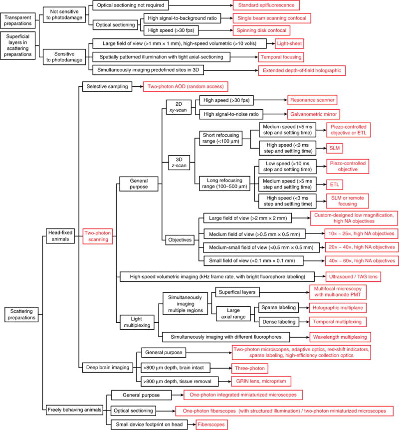
Flowchart to choose imaging platform.
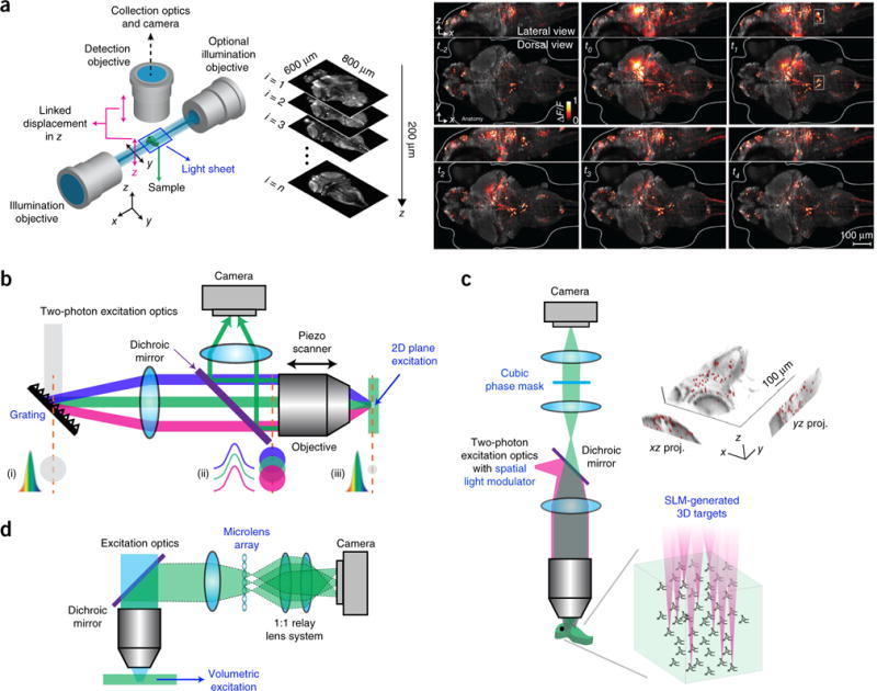
Wide-field imaging. (a) Left, a schematic of a light-sheet microscope. Right, whole-brain neuronal activity of a larval zebrafish recorded with a light-sheet microscope. Brighter hues represent active neurons. Reprinted and adapted from ref. , Macmillan Publishers Limited. (b) Schematic of a microscope using temporal focusing. (i–iii) Temporal and spatial cross-section profiles of the laser beam impinging on the grating (i), at the back aperture (ii) and focal plane of the objective (iii) are shown. Colors indicate different spectrum components. Adapted from ref. , Macmillan Publishers Limited. (c) Holographic microscope with extended depth of field. Right, calcium imaging of 49 neurons targeted simultaneously on a zebrafish. Image reprinted from ref. , Frontiers. (d) Schematic of a light-field microscope. Adapted from ref. , Macmillan Publishers Limited.
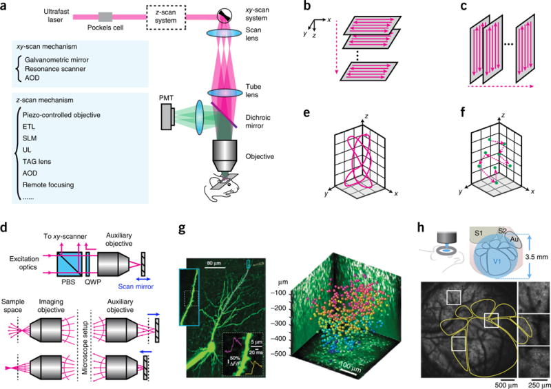
Two-photon microscopy. (a) Optical layout of a two-photon laser scanning microscope. (b) Traditional scan trajectory for volumetric imaging. (c) Scan trajectory enabled by ultrasound or TAG lens. (d) Principle of remote focusing. PBS, polarization beam splitter. QWP, quarter waveplate. Adapted from ref. with permission from the National Academy of Sciences. (e) Custom scanning. Adapted from ref. with permission from the National Academy of Sciences. (f) Random-access scanning in a 3D volume. (g) Examples of data taken with 3D random-access hopping microscope using AODs. Left, calcium imaging of a CA1 pyramidal cell; purple dots represent the scanning points in a dendrite. Right, neurons color coded to depth in mouse visual cortex, imaged with AODs. Reprinted from ref. , Macmillan Publishers Limited. (h) Large FOV microscope. Bottom, calcium imaging of a transgenic mouse expressing GCaMP6s. The yellow outlines indicate the anatomy mapped out by intrinsic signal optical imaging. Reprinted from ref. , Macmillan Publishers Limited.
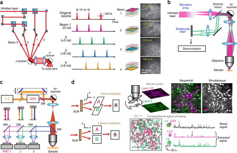
Multiplexing two-photon microscopy. (a) Temporal multiplexing. The single laser beam is split, and one beamlet is delayed such that the pulse trains are interleaved in time. Right, example of a four-plane calcium imaging on mouse brain. Reprinted from ref. , Macmillan Publishers Limited. (b) Multifocal multiphoton microscope. Two beamlets are ray-traced. Adapted from ref. with permission from the Optical Society of America. (c) Wavelength multiplexing. Pulse trains from two lasers (Ti:S, Ti:Sapphire and OPO) target two fluorophores with different two-photon absorption spectra. DM, dichroic mirror. Adapted from ref. , Macmillan Publishers Limited. (d) Holographic simultaneous multiplane imaging. An SLM splits the laser beam to illuminate different axial planes simultaneously. The right panel shows simultaneous calcium imaging on layers 2/3 and 5 on a GCaMP6f-transfected mouse visual cortex in vivo and post hoc signal separation. Reprinted and adapted from ref. with permission from Elsevier.
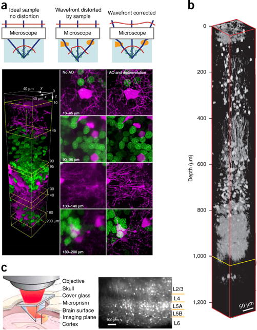
Deep brain imaging. (a) Adaptive optics (AO). Top, a simple model of optical focus formation for different situations. Reprinted from ref. , Macmillan Publishers Limited. Bottom, two-color confocal imaging in a zebrafish optic tectum in vivo with and without adaptive optics. Oligodendrocytes are shown in magenta; neuronal nuclei in green. Reprinted from ref. , Macmillan Publishers Limited. (b) In vivo three-photon imaging of red fluorescence protein labeled neurons in mouse brain. Reprinted from ref. , Macmillan Publishers Limited. (c) Microprism-assisted imaging. Reprinted from ref. with permission from Elsevier.
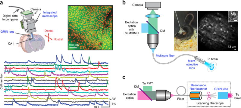
Imaging freely behaving animals. (a) Epifluorescence miniature microscope. Top right, mean fluorescence image from a head-mounted microscope in a behaving GCaMP3-transfected mouse. Identified cells are shown in red. Bottom, fluorescence signals for 15 cells. Reprinted and adapted from ref. , Macmillan Publishers Limited. (b) Epifluorescence fiberscope. Right, a freely behaving mouse with the fiberscope fixed to its skull and calcium imaging of molecular layer interneurons recorded with structured illumination. Inset, single interneuron with a portion of a dendrite (arrow). DM, dichroic mirror. Reprinted and adapted from ref. with permission from Elsevier. (c) Two-photon scanning fiberscope. Adapted from ref. with permission from the Optical Society of America.
References
-
- Crick FHC. Thinking about the brain. Scientific American. 1979;241:219–232. - PubMed
-
- Smetters D, Majewska A, Yuste R. Detecting action potentials in neuronal populations with calcium imaging. Methods. 1999;18:215–221. - PubMed
-
- Ahrens MB, Orger MB, Robson DN, Li JM, Keller PJ. Whole-brain functional imaging at cellular resolution using light-sheet microscopy. Nat Methods. 2013;10:413–420. Example of whole-brain functional imaging in vivo. - PubMed
-
- Denk W, Strickler JH, Webb WW. Two-photon laser scanning fluorescence microscopy. Science. 1990;248:73–76. Invention of two-photon microscopy. - PubMed
-
- Yuste R, Denk W. Dendritic spines as basic functional units of neuronal integration. Nature. 1995;375:682–684. - PubMed
Publication types
MeSH terms
Substances
Grants and funding
LinkOut - more resources
Full Text Sources
Other Literature Sources
