

The Extended-Synaptotagmins
- PMID: 28363589
- PMCID: PMC5642939
- DOI: 10.1016/j.bbamcr.2017.03.013
The Extended-Synaptotagmins
Abstract
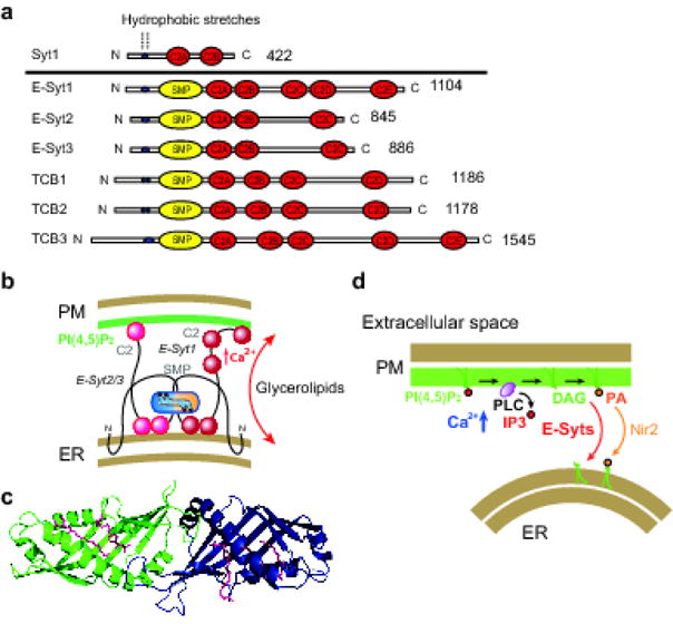
The extended-synaptotagmins (tricalbins in yeast) derive their name from their partial domain structure similarity to the synaptotagmins, which are characterized by an N-terminal membrane anchor and cytosolically exposed C2 domains. However, they differ from the synaptotagmins in localization and function. The synaptotagmins tether secretory vesicles, including synaptic vesicles, to the plasma membrane (PM) via their C2 domains and regulate their Ca2+ triggered exocytosis. In contrast, the extended-synaptotagmins are resident proteins of the endoplasmic reticulum (ER), which tether this organelle to the plasma membrane via their C2 domains, but not as a premise to fusion of the two membranes. They transport glycerolipids between the two bilayers via their lipid-harboring SMP domains and Ca2+ regulates their membrane tethering and lipid transport function. Additionally, the extended-synaptotagmins are more widely expressed in different organisms, as they are present not only in animal cells, but also in fungi and plants, which do not express the synaptotagmins. Thus, they have a more general function than the synaptotagmins, whose appearance in animal species correlated with the occurrence of Ca2+ triggered exocytosis. This article is part of a Special Issue entitled: Membrane Contact Sites edited by Christian Ungermann and Benoit Kornmann.
Keywords: C2; SMP; Synaptotagmin; TULIP; Tricalbin.
Copyright © 2017. Published by Elsevier B.V.
Figures
References
-
- Perin MS, Fried VA, Mignery GA, Jahn R, Sudhof TC. Phospholipid binding by a synaptic vesicle protein homologous to the regulatory region of protein kinase C. Nature. 1990;345:260–263. - PubMed
-
- Brose N, Petrenko AG, Sudhof TC, Jahn R. Synaptotagmin: a calcium sensor on the synaptic vesicle surface. Science. 1992;256:1021–1025. - PubMed
-
- Chapman ER. How does synaptotagmin trigger neurotransmitter release? Annual review of biochemistry. 2008;77:615–641. - PubMed
Publication types
MeSH terms
Substances
Grants and funding
LinkOut - more resources
Full Text Sources
Other Literature Sources
Molecular Biology Databases
Miscellaneous
