. 2017 Jul 15:136:99-108.
doi: 10.1016/j.bcp.2017.03.018. Epub 2017 Mar 29.

Characterization of signal bias at the GLP-1 receptor induced by backbone modification of GLP-1

Affiliations

Characterization of signal bias at the GLP-1 receptor induced by backbone modification of GLP-1

Marlies V Hager et al. Biochem Pharmacol. .

Abstract

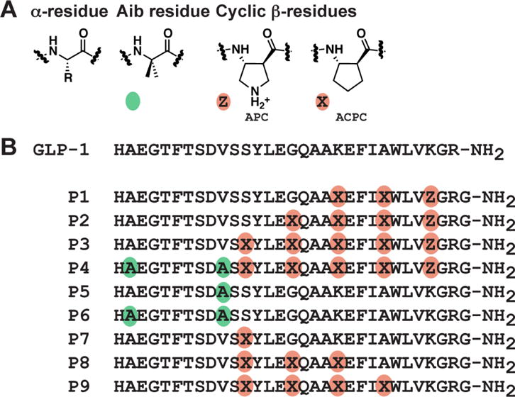
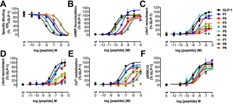
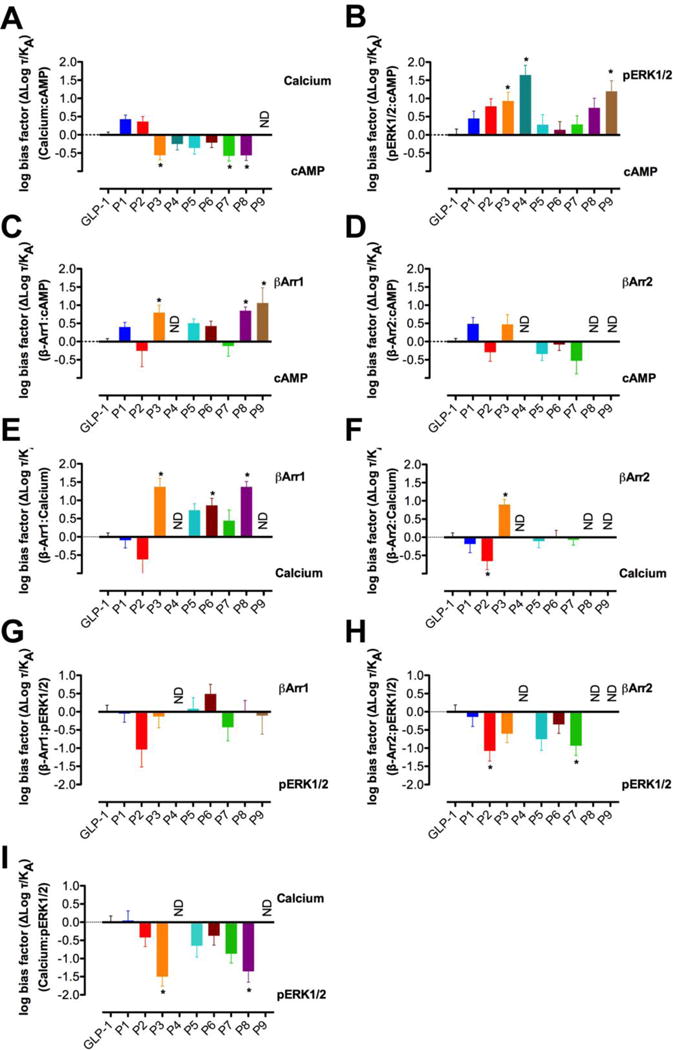
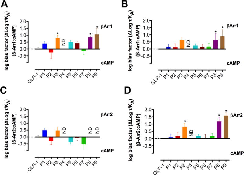
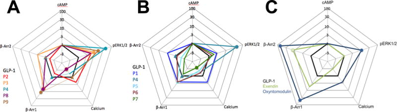
The glucagon-like peptide-1 receptor (GLP-1R) is a class B G protein-coupled receptor that is a major therapeutic target for the treatment of type 2 diabetes. Activation of this receptor promotes insulin secretion and blood glucose regulation. The GLP-1R can initiate signaling through several intracellular pathways upon activation by GLP-1. GLP-1R ligands that preferentially stimulate subsets among the natural signaling pathways ("biased agonists") could be useful as tools for elucidating the consequences of specific pathways and might engender therapeutic agents with tailored effects. Using HEK-293 cells recombinantly expressing human GLP-1R, we have previously reported that backbone modification of GLP-1, via replacement of selected α-amino acid residues with β-amino acid residues, generates GLP-1 analogues with distinctive preferences for promoting G protein activation versus β-arrestin recruitment. Here, we have explored the influence of cell background across these two parameters and expanded our analysis to include affinity and other key signaling pathways (intracellular calcium mobilization and ERK phosphorylation) using recombinant human GLP-1R expressed in a CHO cell background, which has been used extensively to demonstrate biased agonism of GLP-1R ligands. The new data indicate that α/β-peptide analogues of GLP-1 exhibit a range of distinct bias profiles relative to GLP-1 and that broad assessment of signaling endpoints is required to reveal the spectrum of behavior of modified peptides. These results support the view that backbone modification via α→β amino acid replacement can enable rapid discovery of peptide hormone analogues that display substantial signal bias at a cognate GPCR.

Keywords: Biased agonism; Cell signaling; G protein coupled receptor; Glucagon-like peptide-1 receptor; Peptides.


Conflict of interest statement

Conflict of interest statement

The authors declare the following competing financial interest(s): S.H.G. is an inventor on a patent application covering GLP-1 analogues described here; S.H.G. is a cofounder of Longevity Biotech, Inc., which is pursuing biomedical applications of α/β-peptides.

Figures

Figure 1
Figure 1
A. Amino acids used in this study. Colored circles indicate non-natural substitutions: green circles represent the non-proteinogenic α-residue Aib, and orange circles represent ring-constrained β-residues (X = ACPC, Z = APC). B. GLP-1(7–36)NH2 and α/β-peptide analogues 1 - 9 (based on GLP-1(7–37)NH2). Each peptide has a free N-terminus and a primary amide at the C-terminus.
Figure 2
Figure 2
Binding and signaling profiles of GLP-1 and α- and α/β-peptides P1P9 in FlpInCHO cells stably expressing the human GLP-1R. Concentration-response curves for (A) GLP-1R binding, (B) cAMP accumulation, (C) Ca2+ mobilization, (D) ERK1/2 phosphorylation, (E) β-Arrestin-1 recruitment, and (F) β-Arrestin-2 recruitment. Data are normalized to the maximum response elicited by GLP-1 in each assay, and analyzed using a three-parameter logistic equation. Values are the mean + S.E.M. of three to four individual experiments, conducted in duplicate.
Figure 3
Figure 3
Bias factors for α- and α/β-peptides P1P9 relative to GLP-1 in Ca2+ mobilization relative to cAMP accumulation (A), ERK1/2 phosphorylation relative to cAMP accumulation (B), β-Arrestin-1 recruitment relative to cAMP accumulation (C), β-Arrestin-2 recruitment relative to cAMP accumulation (D), β-Arrestin-1 recruitment relative to Ca2+ mobilization (E), β-Arrestin-2 recruitment relative to Ca2+ mobilization (F), β-Arrestin-1 recruitment relative to ERK1/2 phosphorylation (G), and β-Arrestin-2 recruitment relative to ERK1/2 phosphorylation (H). Changes in log (τ/KA) were calculated to provide a measure of the degree of stimulus bias exhibited between different signaling pathways relative to that of the reference agonist GLP-1. Values are the mean ± SEM of three to four individual experiments, conducted in duplicate. * statistically significant difference from GLP-1 using one-way analysis of variance followed by Dunnett’s test.
Figure 4
Figure 4
Comparison of the bias factors for α- and α/β-peptides P1P9 relative to GLP-1 for β-Arrestin-1 recruitment versus cAMP accumulation between FlpInCHO cells (A) and HEK293 cells (B) and for β-Arrestin-2 recruitment versus cAMP accumulation between FlpInCHO cells (C) and HEK293 cells (D). Changes in log (τ/KA) were calculated to provide a measure of the degree of stimulus bias exhibited between different signaling pathways relative to that of the reference agonist GLP-1. * statistically significant difference from GLP-1 using one way analysis of variance followed by Dunnett’s test (P<0.05).
Figure 5
Figure 5
Webs of bias for α- and α/β-peptides P1P9 (A, B) and known biased agonists exendin-4 and oxyntomodulin (C) relative to GLP-1 in FlpInCHO cells stably expressing the human GLP-1R. Circles represent data that are significantly biased. Triangles represent data where no value could be defined. The τ/KA ratio extracted from standard concentration-response data is used to calculate bias factors (ΔΔ(τ/KA) through normalization of the transduction coefficient (τ/KA) to a reference ligand (GLP-1) and reference pathway (cAMP accumulation).

Similar articles

Cited by

References

    1. Zaccardi F, Webb DR, Yates T, Davies MJ. Pathophysiology of type 1 and type 2 diabetes mellitus: a 90-year perspective. Postgrad Med J. 2016;92:63–69. - PubMed
    1. DeFronzo RA. Pathogensis of Type 2 (non-insulin dependent) diabetes mellitus: a balanced overview. Diabetologia. 1992;35:389–397. - PubMed
    1. Hundal RS, Inzucchi SE. Metformin: new understandings, new uses. Drugs. 2003;63:1879–1894. - PubMed
    1. Swinnen SG, Hoekstra JB, DeVries JH. Insulin therapy for type 2 diabetes. Diabetes Care. 2009;32(Suppl. 2):S253–259. - PMC - PubMed
    1. Holst JJ, Knop FK, Vilsboll T, Krarup T, Madsbad S. Loss of incretin effect is a specific, important, and early characteristic of type 2 diabetes. Diabetes Care. 2011;34(Suppl 2):S251–257. - PMC - PubMed

Publication types

Substances