. 2017 May 15;198(10):3857-3868.
doi: 10.4049/jimmunol.1601851. Epub 2017 Mar 31.

CD137 Plays Both Pathogenic and Protective Roles in Type 1 Diabetes Development in NOD Mice

Affiliations

CD137 Plays Both Pathogenic and Protective Roles in Type 1 Diabetes Development in NOD Mice

Matthew H Forsberg et al. J Immunol. .

Abstract

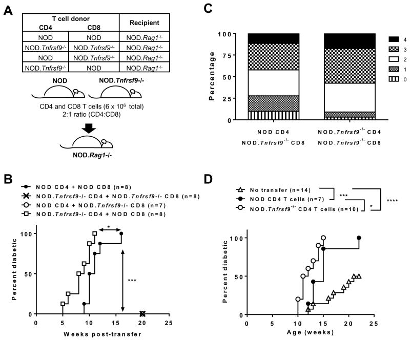
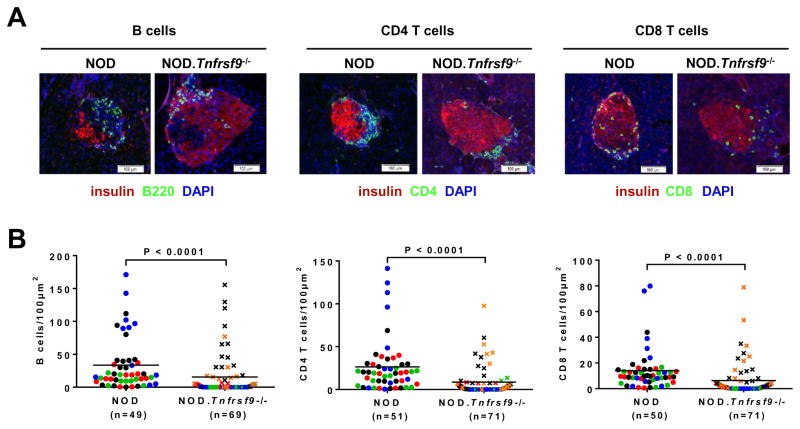
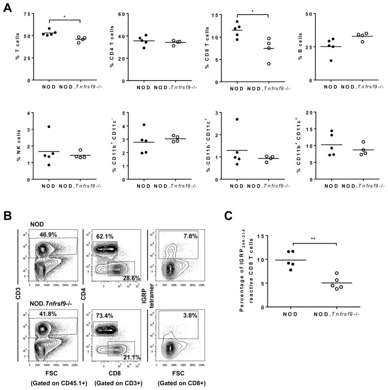
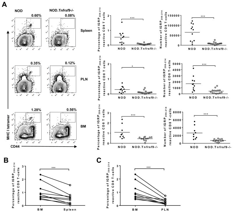
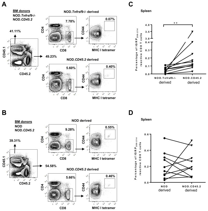
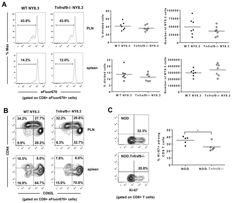
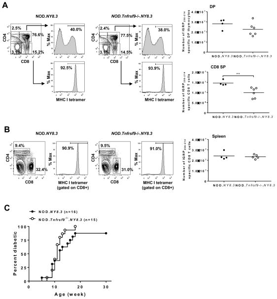
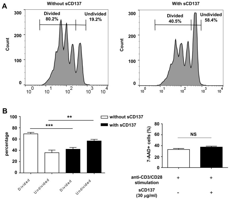
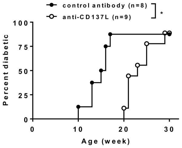
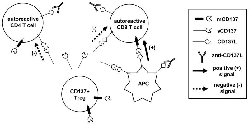
We previously reported that CD137 (encoded by Tnfrsf9) deficiency suppressed type 1 diabetes (T1D) progression in NOD mice. We also demonstrated that soluble CD137 produced by regulatory T cells contributed to their autoimmune-suppressive function in this model. These results suggest that CD137 can either promote or suppress T1D development in NOD mice depending on where it is expressed. In this study, we show that NOD.Tnfrsf9-/- CD8 T cells had significantly reduced diabetogenic capacity, whereas absence of CD137 in non-T and non-B cells had a limited impact on T1D progression. In contrast, NOD.Tnfrsf9-/- CD4 T cells highly promoted T1D development. We further demonstrated that CD137 was important for the accumulation of β cell-autoreactive CD8 T cells but was dispensable for their activation in pancreatic lymph nodes. The frequency of islet-infiltrating CD8 T cells was reduced in NOD.Tnfrsf9-/- mice in part because of their decreased proliferation. Furthermore, CD137 deficiency did not suppress T1D development in NOD mice expressing the transgenic NY8.3 CD8 TCR. This suggests that increased precursor frequency of β cell-autoreactive CD8 T cells in NY8.3 mice obviated a role for CD137 in diabetogenesis. Finally, blocking CD137-CD137 ligand interaction significantly delayed T1D onset in NOD mice. Collectively, our results indicate that one important diabetogenic function of CD137 is to promote the expansion and accumulation of β cell-autoreactive CD8 T cells, and in the absence of CD137 or its interaction with CD137 ligand, T1D progression is suppressed.


Figures

Figure 1
Figure 1. CD137 expression in CD4 and CD8 T cells differentially controls their diabetogenic activity
(A) Schematic diagram showing the experimental design of the CD8 and CD4 T cell co-transfer into NOD.Rag1−/− recipients. (B) T1D incidence study of NOD.Rag1−/− recipients depicted in Figure 1A. CD4 and CD8 T cells isolated from 6–8 week old female NOD and/or NOD.Tnfrsf9−/− mice were injected into 6–8 week old NOD.Rag1−/− females. Recipients were analyzed for the development of T1D weekly over the course of 20 weeks post-transfer. (C) Histological analysis of NOD.Rag1−/− recipients in Figure 1B that did not progress to T1D at the end of incidence study. Islets were scored 0 to 4 based on the severity of insulitis: 0, no lesions; 1, peri-insular leukocytic aggregates, 2, <25% islet destruction; 3, >25% islet destruction; and 4, complete islet destruction. The result is presented as the relative proportion of each score. More than 200 islets were scored in each recipient group. (D) T1D incidence study of NOD.Rag1−/−.NY8.3 female recipients of 3x10⁶ CD4 T cells from 6–8 week old NOD and NOD.Tnfrsf9−/− female mice. Recipients were infused with purified CD4 T cells at 6–8 weeks old and followed for the development of T1D every week over the course of 22 weeks post transfer. *P < 0.05, ***P < 0.005, ***P < 0.001 by Log-rank test.
Figure 2
Figure 2. Accumulation of B cells, CD4 T cells, and CD8 T cells in islets is reduced in the NOD.Tnfrsf9−/− strain compared to NOD mice
Pancreata from 10-week-old NOD and NOD.Tnfrsf9−/− females were processed for immunofluorescence staining to determine the numbers of islet infiltrating B cells, CD4 T cells, and CD8 T cells as described in Materials and Methods. (A) Representative immunofluorescence staining of islets from NOD and NOD.Tnfrsf9−/− female mice (B220, CD4, and CD8 = green; insulin = red; DAPI = blue). (B) Numbers of B cells, CD4 T cells, or CD8 T cells per 100μm2 of islet area. Each symbol represents one islet. Islets from the same mouse are color coded. Results are pooled from 4 NOD and 5 NOD.Tnfrsf9−/− mice. Statistical analysis was carried out by Mann-Whitney test.
Figure 3
Figure 3. Intra-islet accumulation of CD8 T cells is proportionally reduced in the NOD.Tnfrsf9−/− strain compared to NOD mice
(A) Flow cytometric analysis of various islet infiltrating leukocyte populations. Islets were isolated from 9–12 week old NOD and NOD.Tnfrsf9−/− female mice and dissociated into single cell suspension. Leukocytes were identified by CD45.1 expression and the frequencies of different cell populations, including total T cells (CD3+), CD4 T cells (CD3+ CD4+), CD8 T cells (CD3+ CD8+), B cells (CD19+), NK cells (CD3 DX5+), and myeloid cells (CD11b+ CD11c, CD11b+ CD11c+, and CD11b CD11c+) were calculated as percentages of total CD45.1+ cells. Each symbol represents cells pooled from 2–7 mice. *P < 0.05 by Mann-Whitney test. (B and C) The frequencies of islet associated IGRP206-214 reactive CD8 T cells in NOD and NOD.Tnfrsf9−/− female mice by MHC class I tetramer staining. Islet cells pooled from 2–5 mice (10–12 weeks old) were stained with antibodies against CD45.1, CD3, CD4, and CD8 as well as the IGRP206-214 MHC I tetramer. (B) One representative experiment is shown. (C) Summarized results from five independent experiments. **P < 0.01 by Mann-Whitney test.
Figure 4
Figure 4. The frequencies and numbers of IGRP206-214 reactive CD8 T cells in the spleen, PLN, and BM are significantly lower in NOD.Tnfrsf9−/− than in NOD mice
(A) Cells were isolated from the spleen, PLN, and BM, and strained with antibodies against CD8 and CD44 as well as IGRP206-214 loaded MHC class I tetramers. Representative flow cytometry profiles are shown in the left panels. The percentages and numbers of IGRP206-214 MHC class I tetramer staining of the spleen, PLN, and BM from 10–14 week old NOD and NOD.Tnfrsf9−/− female mice from 4 independent experiments are summarized respectively in the middle and right panels. In one experiment, BM from two mice of each strain were not analyzed. *P < 0.05, ***P < 0.005 by Mann-Whitney test. (B and C) The frequency of IGRP206-214 reactive CD8 T cells is higher in the BM than in the spleen (B) and PLN (C). Paired analysis of IGRP206-214 MHC class I tetramer staining for BM versus spleen and BM versus PLN in NOD mice. ***P < 0.005 by Wilcoxon matched-pairs signed rank test.
Figure 5
Figure 5. CD137 intrinsically promotes the accumulation of IGRP206-214 reactive CD8 T cells
Lethally irradiated (NOD x NOD.CD45.2)F1 mice were infused with equal number of T cell depleted BM cells (2.5x10⁶ per donor strain) from NOD.CD45.2 and NOD.Tnfrsf9−/− (A and C) or NOD.CD45.2 and NOD mice (B and D). The frequencies of CD44high IGRP206-214 reactive CD8 T cells were analyzed in the spleens of the F1 recipients by MHC I tetramer staining at 10–12 weeks post BM reconstitution. The origin of the splenocytes was determined by CD45.1 and CD45.2 expression. The gating strategy is shown in panels A and B. The summarized results of two independent experiments are shown in panels C and D. **P < 0.01 by Wilcoxon matched-pairs signed rank test.
Figure 6
Figure 6. Regulation of autoreactive CD8 T cell proliferation by CD137
(A and B) CD137 is not required for initial activation of β-cell autoreactive CD8 T cells in PLNs. CD8 T cells purified from NOD.NY8.3 and NOD.Tnfrsf9−/−.NY8.3 mice were labelled with eFluor670 cell proliferation dye and transferred into NOD recipients. Six days later, proliferation of adoptively transferred NY8.3 CD8 T cells was analyzed in the spleen and PLN. (A) Representative flow cytometry profiles of cell division are shown on the left. The results of cell proliferation (middle panels) and the recovered numbers (right panels) of adoptively transferred NY8.3 CD8 T cells are summarized from two independent experiments. Each symbol represents one recipient mouse. (B) Representative flow cytometry profiles of CD44 and CD62L expression on the adoptively transferred NY8.3 CD8 T cells. (C) Intra-islet proliferation of CD8 T cells is reduced in the NOD.Tnfrsf9−/− strain compared to the NOD mice. Ki-67 expression was analyzed by flow cytometry in islet infiltrating CD8 T cells from 10–14 week-old NOD and NOD.Tnfrsf9−/− female mice. Representative flow cytometry profiles of the percentages of Ki-67+ CD8 T cells are shown (left). The results are summarized from 5 independent experiments (right). Each symbol represents islet cells pooled from 3–5 mice. *P < 0.05 by Mann-Whitney test.
Figure 7
Figure 7. CD137 deficiency does not alter diabetes development in NOD mice transgenically expressing TCRs of the NY8.3 clonal type
(A) Thymi from 6–7 week-old NOD.NY8.3 and NOD.Tnfrsf9−/−.NY8.3 female mice were analyzed for IGRP206-214 specific double-positive (DP) and CD8 single-positive (SP) thymocytes. Representative flow cytometry profiles for each strain are shown (left and middle panels). The results are summarized in the right panels. Each symbol represents one mouse. **P < 0.01 by Mann-Whitney test. (B) Spleens from 6–7 week-old NOD.NY8.3 and NOD.Tnfrsf9−/−.NY8.3 female mice were analyzed for IGRP206-214 specific CD8 T cells. Representative flow cytometry profiles for each strain are shown (left and middle panels). The results are summarized in the right panels. Each symbol represents one mouse. (C) T1D incidence study of NOD.NY8.3 and NOD.Tnfrsf9−/−.NY8.3 female mice. T1D development was monitored weekly using urine glucose strips with onset defined by two consecutive readings of > 250 mg/dl. Diabetes development is not significantly different between NOD.NY8.3 and NOD.Tnfrsf9−/−.NY8.3 mice.
Figure 8
Figure 8. Soluble CD137 suppresses CD8 T cell activation
CD8 T cells purified from 8–10 week-old female NOD mice were labeled by CFSE (final concentration: 0.5 uM), stimulated by 25,000 CD3/CD28 dynabeads and cultured with or without recombinant soluble CD137 (30 ug/ml) on 96-well u-bottom culture plate. After 3 days of culture, CFSE-dilution and 7-AAD positive population was quantified on FACS Calibur. (A) One representative of four experiments for CFSE dilution without soluble CD137 (left) and with soluble CD137 (right) is shown. (B) Soluble CD137 significantly increases the number of undivided CD8 T cells and decreases the number of divided CD8 T cells. Mean percentage of divided and undivided cells is shown (n = 4 experiments). (C) 7-AAD staining shows no difference in CD8 cell death after 3 days of culture with or without soluble CD137 (mean of n = 4 experiments). **P < 0.01; ***P < 0.005 by unpaired t test. sCD137: soluble CD137. NS: not significant.
Figure 9
Figure 9. CD137L blockage delays T1D onset in NOD mice
Starting at 9 weeks of age, NOD female mice were injected every other week for a total of 5 injections with 250μg of anti-CD137L (TKS-1) each treatment or an isotype control antibody. T1D development was monitored weekly using urine glucose strips with onset defined by two consecutive readings of > 250 mg/dl. *P < 0.05 by Log-rank test.
Figure 10
Figure 10. Schematic diagram illustrating the functions of CD137-CD137L interactions in the regulation of β-cell autoreactive T cells
Treg-derived soluble CD137 (sCD137) binds to CD137L expressed on T cells and suppresses their activation. CD137L expressed by APC binds to membrane CD137 (mCD137) on β-cell autoreactive CD8 T cells to promote their expansion and accumulation. For therapeutic purposes, recombinant sCD137 can suppress activation of β-cell autoreactive T cells through direct binding to CD137L expressed on their surface as well as by blocking the interaction between mCD137 on CD8 T cells and CD137L on APC. CD137L blocking antibody inhibits interaction between mCD137 on CD8 T cells and CD137L on APC. CD137L blocking antibody also binds to CD137L on T cells without inducing an inhibitory effect but it prevents the suppressive function of sCD137.

Similar articles

Cited by

References

    1. Watts TH. TNF/TNFR family members in costimulation of T cell responses. Annu Rev Immunol. 2005;23:23–68. - PubMed
    1. Arch RH, Thompson CB. 4-1BB and Ox40 are members of a tumor necrosis factor (TNF)-nerve growth factor receptor subfamily that bind TNF receptor-associated factors and activate nuclear factor kappaB. Mol Cell Biol. 1998;18:558–565. - PMC - PubMed
    1. Jang IK, Lee ZH, Kim YJ, Kim SH, Kwon BS. Human 4-1BB (CD137) signals are mediated by TRAF2 and activate nuclear factor-kappa B. Biochemical and biophysical research communications. 1998;242:613–620. - PubMed
    1. Cannons JL, Hoeflich KP, Woodgett JR, Watts TH. Role of the stress kinase pathway in signaling via the T cell costimulatory receptor 4-1BB. Journal of immunology. 1999;163:2990–2998. - PubMed
    1. Cannons JL, Choi Y, Watts TH. Role of TNF receptor-associated factor 2 and p38 mitogen-activated protein kinase activation during 4-1BB-dependent immune response. Journal of immunology. 2000;165:6193–6204. - PubMed

Publication types

MeSH terms

Substances