. 2017 May 15;198(10):3835-3845.
doi: 10.4049/jimmunol.1602122. Epub 2017 Mar 31.

Varied Role of Ubiquitylation in Generating MHC Class I Peptide Ligands

Affiliations

Varied Role of Ubiquitylation in Generating MHC Class I Peptide Ligands

Jiajie Wei et al. J Immunol. .

Abstract

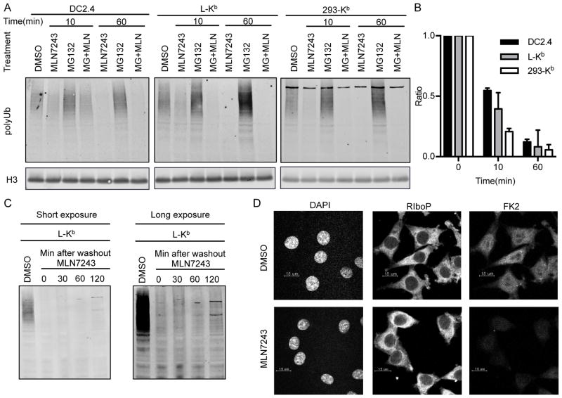
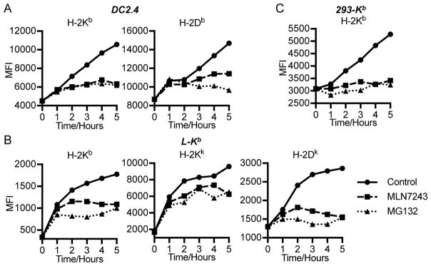
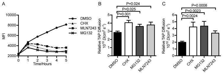
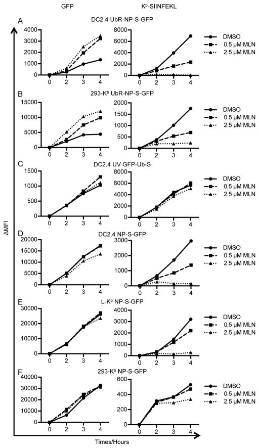
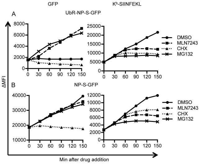
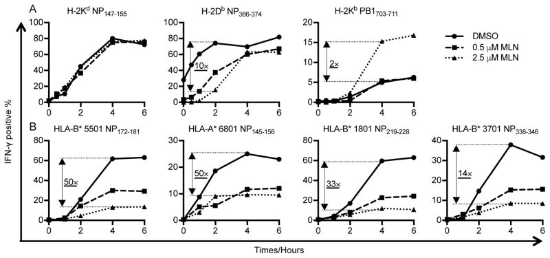
CD8+ T cell immunosurveillance is based on recognizing oligopeptides presented by MHC class I molecules. Despite decades of study, the importance of protein ubiquitylation to peptide generation remains uncertain. In this study, we examined the ability of MLN7243, a recently described ubiquitin-activating enzyme E1 inhibitor, to block overall cytosolic peptide generation and generation of specific peptides from vaccinia- and influenza A virus-encoded proteins. We show that MLN7243 rapidly inhibits ubiquitylation in a variety of cell lines and can profoundly reduce the generation of cytosolic peptides. Kinetic analysis of specific peptide generation reveals that ubiquitylation of defective ribosomal products is rate limiting in generating class I peptide complexes. More generally, our findings demonstrate that the requirement for ubiquitylation in MHC class I-restricted Ag processing varies with class I allomorph, cell type, source protein, and peptide context. Thus, ubiquitin-dependent and -independent pathways robustly contribute to MHC class I-based immunosurveillance.


Figures

Figure 1
Figure 1. MLN7243 blocks protein ubiquitylation
(A) DC2.4, L-Kb and 293-Kb cells were treated with 2.5 μM MLN7243, 10 μM MG132, or the combination of these two drugs for 10 or 60 min. Whole cell lysates were blotted with FK1 to detect polyubiquitylated proteins. Histone-H3 was used as the loading control. (B) Median intensity of each band was used for quantification. Results from three independent experiments are shown. (C) L-Kb cells were treated with 2.5 μM MLN7243, washed three times with PBS, and cultured in medium without MLN7243 for the indicated times. Whole cell lysates were blotted with FK1 to detect polyubiquitylated proteins. (D) L-Kb cells were cultured in the presence or absence of 2.5 μM MLN7243 for 1 h. DAPI, anti-ribosomal protein P antibody and FK2 were used in immunofluorescence microscopy to detect nuclei (left panel), ribosomes (middle panel) and ubiquitylated proteins (right panel), respectively.
Figure 2
Figure 2. MLN7243 inhibits MHC class I-restricted endogenous antigen surface expression
DC2.4 (A), L-Kb (B), and 293-Kb (C) cells were washed for 2 min in cold citric acid buffer to destroy native cell surface MHC-I complexes. Cells were then cultured in the presence of 2.5 μM MLN7243 or 10 μM MG132 or equal amount of DMSO, harvested at indicated time points, and analyzed for surface MHC-I complexes by flow cytometry. MFI: medium fluorescent intensity.
Figure 3
Figure 3. MLN7243 inhibits cytosolic peptide generation
(A) TAP1-GFP cells were washed for 2 min in cold citric acid buffer, then cultured in the presence of DMSO, cycloheximide (CHX), MLN7243 or MG132, harvested at indicated time points, and analyzed for surface MHC-I complexes by flow cytometry. (B) Lateral mobility of TAP in TAP1-GFP cells determined by FRAP assay. (C) Diffusion rate of TAP1-GFP measured by fluorescence correlation spectroscopy. GraphPad Prism software was used to calculate P values (Unpaired t test).
Figure 4
Figure 4. MLN7243 selectively inhibits Kb-SIINFEKL presentation from rVV-expressed proteins
DC2.4 (A, C and D), L-Kb (E), and 293-Kb (B and F) cells were infected for 1 h with rVV expressing UbR-NP-S-GFP (A and B), partially UV-inactivated rVV expressing GFP-Ub-S (C), or rVV expressing NP-S-GFP (D–F). Cells were then cultured in the presence of DMSO, 0.5 μM MLN7243, or 2.5 μM MLN7243 and harvested at indicated time points. Levels of GFP (left panels) and surface Kb-SIINFEKL (right panels) were determined by flow cytometry. ΔMFI: medium fluorescent intensity after background subtraction.
Figure 5
Figure 5. Fine kinetics of inhibitor blockade
L-Kb cells were infected with rVVs expressing UbR-NP-S-GFP (A) or NP-S-GFP (B) for 1 h. At 3 h, DMSO, MLN7243, CHX or MG132 were added into cell cultures. Cells were harvested at indicated time points after drug addition. Levels of GFP (left panels) and surface Kb-SIINFEKL (right panels) were determined by flow cytometry. ΔMFI: medium fluorescent intensity after background subtraction.
Figure 6
Figure 6. MLN7243 differentially affects mouse and human MHC-I presentation of IAV antigens
Antigen presenting cells were infected with IAV PR8 for 60 min, cultured in the presence of DMSO, 0.5 μM MLN7243, or 2.5 μM MLN7243, washed, and used to stimulate T cell lines with BFA addition at indicated time points. Antigen presentation of mouse H-2Kd restricted NP147–155, H-2Db restricted NP366–374 and H-2Kb restricted PB1703–711 (A) and human HLA-B* 5501 restricted NP172–181, HLA-A* 6801 restricted NP145–156, HLA-B* 3701 NP338–346 and HLA-B* 1801 restricted NP219–228 (B) was assessed by specific TCD8+ cultures by IFN-γ ICS.

Similar articles

Cited by

References

    1. Anton LC, Yewdell JW. Translating DRiPs: MHC class I immunosurveillance of pathogens and tumors. J Leukoc Biol. 2014;95:551–562. - PMC - PubMed
    1. Bourdetsky D, Schmelzer CE, Admon A. The nature and extent of contributions by defective ribosome products to the HLA peptidome. Proc Natl Acad Sci USA. 2014;111:E1591–1599. - PMC - PubMed
    1. Purcell AW, Croft NP, Tscharke DC. Immunology by numbers: quantitation of antigen presentation completes the quantitative milieu of systems immunology! Curr Opin Immunol. 2016;40:88–95. - PubMed
    1. Zanker D, Waithman J, Yewdell JW, Chen W. Mixed Proteasomes Function To Increase Viral Peptide Diversity and Broaden Antiviral CD8+ T Cell Responses. J Immunol. 2013;191:52–59. - PMC - PubMed
    1. Croft NP, Smith SA, Wong YC, Tan CT, Dudek NL, Flesch IEA, Lin LCW, Tscharke DC, Purcell AW. Kinetics of Antigen Expression and Epitope Presentation during Virus Infection. PLoS Pathog. 2013;9:e1003129. - PMC - PubMed

MeSH terms