. 2017 May 17;83(11):e00175-17.
doi: 10.1128/AEM.00175-17. Print 2017 Jun 1.

Mechanisms for Pseudoalteromonas piscicida-Induced Killing of Vibrios and Other Bacterial Pathogens

Affiliations

Mechanisms for Pseudoalteromonas piscicida-Induced Killing of Vibrios and Other Bacterial Pathogens

Gary P Richards et al. Appl Environ Microbiol. .

Abstract

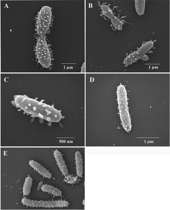
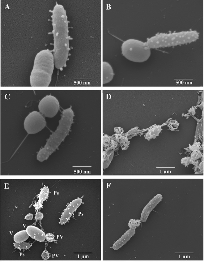
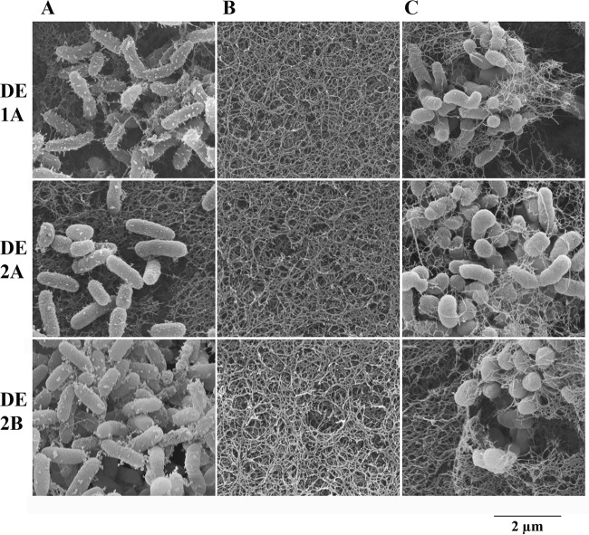
Pseudoalteromonas piscicida is a Gram-negative gammaproteobacterium found in the marine environment. Three strains of pigmented P. piscicida were isolated from seawater and partially characterized by inhibition studies, electron microscopy, and analysis for proteolytic enzymes. Growth inhibition and death occurred around colonies of P. piscicida on lawns of the naturally occurring marine pathogens Vibrio vulnificus, Vibrio parahaemolyticus, Vibrio cholerae, Photobacterium damselae, and Shewanella algae Inhibition also occurred on lawns of Staphylococcus aureus but not on Escherichia coli O157:H7 or Salmonella enterica serovar Typhimurium. Inhibition was not pH associated, but it may have been related to the secretion of a cysteine protease with strong activity, as detected with a synthetic fluorogenic substrate. This diffusible enzyme was secreted from all three P. piscicida strains. Direct overlay of the Pseudoalteromonas colonies with synthetic fluorogenic substrates demonstrated the activity of two aminopeptidase Bs, a trypsin-like serine protease, and enzymes reactive against substrates for cathepsin G-like and caspase 1-like proteases. In seawater cultures, scanning electron microscopy revealed numerous vesicles tethered to the outer surface of P. piscicida and a novel mechanism of direct transfer of these vesicles to V. parahaemolyticus Vesicles digested holes in V. parahaemolyticus cells, while the P. piscicida congregated around the vibrios in a predatory fashion. This transfer of vesicles and vesicle-associated digestion of holes were not observed in other bacteria, suggesting that vesicle binding may be mediated by host-specific receptors. In conclusion, we show two mechanisms by which P. piscicida inhibits and/or kills competing bacteria, involving the secretion of antimicrobial substances and the direct transfer of digestive vesicles to competing bacteria.IMPORTANCEPseudoalteromonas species are widespread in nature and reduce competing microflora by the production of antimicrobial compounds. We isolated three strains of P. piscicida and characterized secreted and cell-associated proteolytic enzymes, which may have antimicrobial properties. We identified a second method by which P. piscicida kills V. parahaemolyticus It involves the direct transfer of apparently lytic vesicles from the surface of the Pseudoalteromonas strains to the surface of Vibrio cells, with subsequent digestion of holes in the Vibrio cell walls. Enzymes associated with these vesicles are likely responsible for the digestion of holes in the cell walls. Pseudoalteromonas piscicida has potential applications in aquaculture and food safety, in control of the formation of biofilms in the environment, and in food processing. These findings may facilitate the probiotic use of P. piscicida to inactivate pathogens and may lead to the isolation of enzymes and other antimicrobial compounds of pharmacological value.

Keywords: Pseudoalteromonas piscicida; Vibrio cholerae; Vibrio parahaemolyticus; Vibrio vulnificus; bacterial inhibition; enzymes; lytic vesicles; predatory bacteria; probiotics; proteases.


Figures

FIG 1
FIG 1
Evolutionary relationships among Pseudoalteromonas piscicida strains and related species based on 16S rRNA gene sequence. The evolutionary relationships were inferred using the neighbor-joining method. The optimal tree with the sum of branch lengths of 0.10271041 is shown. The percentages of replicate trees in which the associated taxa clustered together in the bootstrap test (1,000 replicates) are shown next to the branches. The tree is drawn to scale, with branch lengths in the same units as those of the evolutionary distances used to infer the phylogenetic tree. The evolutionary distances were computed using the Kimura 2-parameter method and are in the units of the number of base substitutions per site. The rate variation among sites was modeled with a gamma distribution (shape parameter = 5). The analysis involved 30 nucleotide sequences. All positions containing gaps and missing data were eliminated. There were a total of 1,155 positions in the final data set. Evolutionary analyses were conducted in MEGA7.
FIG 2
FIG 2
Scanning electron micrographs of P. piscicida strains. (A and B) Multiple morphologies of P. piscicida strain DE1-A with notable vesicles. (C) Large flat vesicles extending outward from the surface of P. piscicida strain DE2-A. (D and E) Typical appearance of P. piscicida strain DE2-B with widely varied lengths.
FIG 3
FIG 3
Scanning electron micrographs of P. piscicida strains in coculture with V. parahaemolyticus O3:K6. (A to C) Apparent transfer of digestive vesicles from P. piscicida to the surfaces of V. parahaemolyticus in P. piscicida strain DE2-A (A and B) and strain DE1-A (C). (D) Vibrios containing vesicle-associated holes, except in center of micrograph, where an intact V. parahaemolyticus appears with vesicles on its surface. (E) P. piscicida (Ps), V. parahaemolyticus (V), and late-stage V. parahaemolyticus with vesicle-digested holes visible (permeabilized vibrios [PV]). (F) Two P. piscicida DE2-A bacteria suspected to be feeding (grazing) on nutrients released by a permeabilized V. parahaemolyticus.
FIG 4
FIG 4
High magnification of P. piscicida strain DE2-C in a V. parahaemolyticus coculture showing large round bulbous vesicles and apparent scars on its surface, suspected to be from the transfer of vesicles to the vibrios. Scale bar signifies 500 nm.
FIG 5
FIG 5
Scanning electron micrographs of agar plugs excised from inhibition assays. Plates containing V. vulnificus in the agar were stabbed with three strains of P. piscicida and incubated at 22°C for 24 h until Pseudoalteromonas colonies and zones of clearing around the colonies became visible. The entire area (colonies, zones of clearing around the colonies, and the area just outside the clear zones) was excised, processed, and subjected to SEM. (A) The outer edges of three colonies of P. piscicida (strains DE1-A, DE2-A, and DE2-B) showing typical morphologies, including extracellular vesicles. (B) Areas within the zone of Vibrio inhibition showing the absence of bacteria. (C) Normal-appearing lawns of V. vulnificus just outside the zone of inhibition. Images taken at ×50,000 magnification; scale bar beneath images = 2.0 μm and applies to all micrographs.
FIG 6
FIG 6
Colonies and fluorogenic assays for enzyme activity. (A) Photographs of colonies of P. piscicida strain DE2-A, zones of clearing around the colonies, and surrounding healthy lawns of V. vulnificus strain MLT362. (B) Same areas as in panel A were overlaid with cellulose acetate membranes containing synthetic fluorogenic (7-amino-4-trifluoromethylcoumarin-linked [AFC]) substrates and photographed under UV illumination at 364 nm to identify proteolytic activity (light areas). Representative results are shown using substrates AFC-002 (l-Arg-AFC), AFC-008 (l-Lys-AFC), AFC-059 (d-Pro-Phe-Arg-AFC), AFC-096 (MeoSuc-Ala-Ala-Pro-Met-AFC), and AFC-120 (N-acetyl-Tyr-Val-Ala-Asp-AFC).

References

    1. Hayashida-Soiza G, Uchida A, Mori N, Kuwahara Y, Ishida Y. 2008. Purification and characterization of antibacterial substances produced by a marine bacterium Pseudoalteromonas haloplanktis strain. J Appl Microbiol 105:1672–1677. doi: 10.1111/j.1365-2672.2008.03878.x. - DOI - PubMed
    1. Yu M, Wang J, Tang K, Shi X, Wang S, Zhu W-M, Zhang X-H. 2012. Purification and characterization of antibacterial compounds of Pseudoalteromonas flavipulchra JG1. Microbiology 158:835–842. doi: 10.1099/mic.0.055970-0. - DOI - PubMed
    1. Isnansetyo A, Kamei Y. 2003. MC21-A, a bactericidal antibiotic produced by a new marine bacterium, Pseudoalteromonas phenolica sp. nov. O-BC30(T), against methicillin-resistant Staphylococcus aureus. Antimicrob Agents Chemother 47:480–488. doi: 10.1128/AAC.47.2.480-488.2003. - DOI - PMC - PubMed
    1. Offret C, Desriac F, Le Chevalier P, Mounier J, Jégou C, Fleury Y. 2016. Spotlight on antimicrobial metabolites from the marine bacteria Pseudoalteromonas: chemodiversity and ecological significance. Mar Drugs 14:129. doi: 10.3390/md14070129. - DOI - PMC - PubMed
    1. Vynne NG, Må M, Nielsen KF, Gram L. 2011. Bioactivity, chemical profiling, and 16S rRNA-based phylogeny of Pseudoalteromonas strains collected on a global research cruise. Mar Biotechnol 13:1062–1073. doi: 10.1007/s10126-011-9369-4. - DOI - PubMed

MeSH terms

LinkOut - more resources