. 2017 Jun;206(2):775-784.
doi: 10.1534/genetics.116.199687. Epub 2017 Mar 31.

Facilitating Neuron-Specific Genetic Manipulations in Drosophila melanogaster Using a Split GAL4 Repressor

Affiliations

Facilitating Neuron-Specific Genetic Manipulations in Drosophila melanogaster Using a Split GAL4 Repressor

Michael-John Dolan et al. Genetics. 2017 Jun.

Abstract

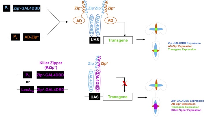
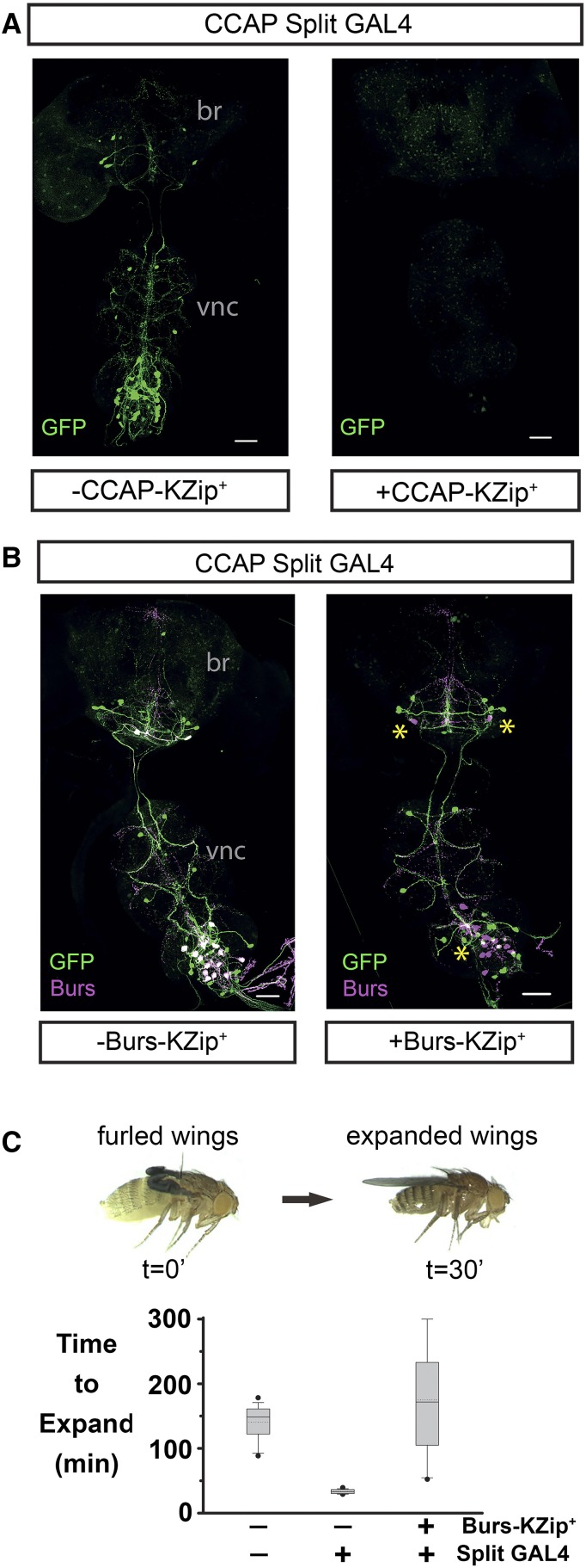
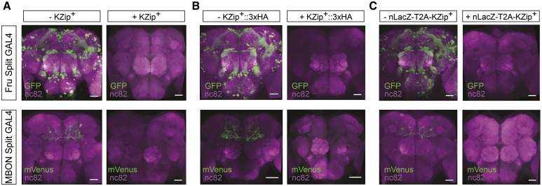
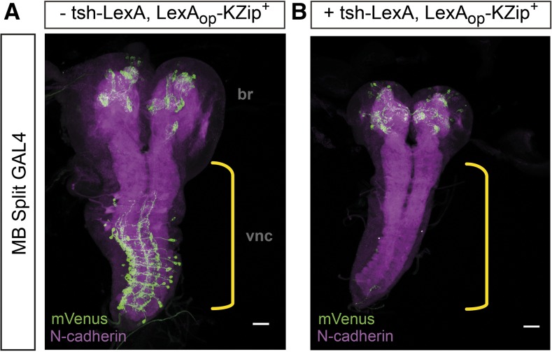
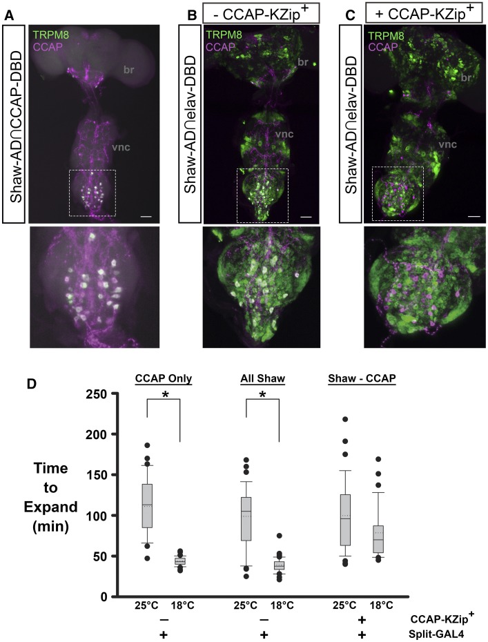
Efforts to map neural circuits have been galvanized by the development of genetic technologies that permit the manipulation of targeted sets of neurons in the brains of freely behaving animals. The success of these efforts relies on the experimenter's ability to target arbitrarily small subsets of neurons for manipulation, but such specificity of targeting cannot routinely be achieved using existing methods. In Drosophila melanogaster, a widely-used technique for refined cell type-specific manipulation is the Split GAL4 system, which augments the targeting specificity of the binary GAL4-UAS (Upstream Activating Sequence) system by making GAL4 transcriptional activity contingent upon two enhancers, rather than one. To permit more refined targeting, we introduce here the "Killer Zipper" (KZip+), a suppressor that makes Split GAL4 targeting contingent upon a third enhancer. KZip+ acts by disrupting both the formation and activity of Split GAL4 heterodimers, and we show how this added layer of control can be used to selectively remove unwanted cells from a Split GAL4 expression pattern or to subtract neurons of interest from a pattern to determine their requirement in generating a given phenotype. To facilitate application of the KZip+ technology, we have developed a versatile set of LexAop-KZip+ fly lines that can be used directly with the large number of LexA driver lines with known expression patterns. KZip+ significantly sharpens the precision of neuronal genetic control available in Drosophila and may be extended to other organisms where Split GAL4-like systems are used.

Keywords: Drosophila; Gal4-UAS; LexA-LexAop; neural circuits; transgene expression.


Figures

Figure 1
Figure 1
Mechanism of Killer Zipper (KZip+) suppression of Split GAL4 activity. (A) The Split GAL4 system consists of functionally distinct transcriptional components, a GAL4 DNA-binding domain (GAL4DBD), and a transcription activation domain (AD), each fused to a heterodimerizing leucine zipper (Zip or Zip+). Each component can be placed under the control of a different promoter (P1 or P2) resulting in two hemidriver lines, which drive the transcriptional components in different populations of cells. When the hemidrivers are combined, Zip+ and Zip dimerize in cells expressing both components, producing a functional Split GAL4 transcription factor capable of transcribing Upstream Activating Sequence (UAS)-transgenes. (B) KZip+ (Zip+-GAL4DBD) consists of the GAL4DBD fragment fused to the Zip+ leucine zipper and can be expressed either directly under the control of a third promoter (P3) or indirectly under such control by a LexA driver (LexAop). In cells that express the Split GAL4 components as well as KZip+, the latter molecule can form homodimers with the Zip-GAL4DBD and thus titrate GAL4DBD partners for the Zip+-AD. Furthermore, the homodimers can compete for binding to UAS sites and block transcription by residual functional Split GAL4 heterodimers.
Figure 2
Figure 2
The KZip+ construct robustly suppresses Split GAL4-mediated transgene expression. (A) Left: Confocal projection view of a wholemount adult CNS showing the CCAP-expressing neurons visualized by a UAS-2xEGFP reporter (green) driven by a Split GAL4 driver (CCAP-GAL4DBD∩elav-VP16AD). Right: Reporter expression is substantially suppressed when the KZip+ is coexpressed in the CCAP neurons. Each image is a representative of n = 8 brains per genotype (the genotypes used in the experiments shown in all figures are listed in File S1; here, and in all subsequent figures, UAS-reporter expression has been amplified by anti-GFP immunostaining to stringently test for suppression of Split GAL4 activity by KZip+). (B) Left: CCAP neurons expressing 2xEGFP (green) as in (A), but double-labeled with anti-Burs antibody (magenta; double-labeled neurons appear white). Right: Expression of KZip+ in the subset of CCAP neurons that coexpresses the burs gene selectively blocks 2xEGFP expression in these neurons, which now label only in magenta (asterisks). Bar, 50 μm for all images in (A) and (B). Each image is a representative of n = 14 brains per genotype. (C) Top: the wings of newly emerged flies are initially furled and become expanded upon execution of a behavioral program governed by the hormone bursicon. The bursicon-induced program is usually executed within the first 30 min of emergence, but is substantially delayed if flies are confined. Confined flies, however, also expand quickly if the complement of CCAP-expressing neurons that express bursicon is artificially stimulated by activation of the cold-sensitive TRPM8 cation channel. Bottom: Box plots indicate the expansion times of control flies that lack the CCAP-AD hemidriver (left), or flies expressing UAS-TRPM8 in the CCAP-expressing neurons under the control of Split GAL4 (CCAP-AD∩CCAP-DBD > UAS-TRPM8) either with (right) or without (middle) KZip+ coexpression in the bursicon neurons. All flies were subjected to a 15 min temperature shift to 18° to activate TRPM8. KZip+ expression in the bursicon-expressing neurons prevents their expression of UAS-TRPM8 and therefore their activation by temperature shift. br, brain; Burs, bursicon promoter; CCAP, Crustacean Cardioactive Peptide; EGFP, enhanced GFP; KZip+, Killer Zipper; UAS, Upstream Activating Sequence; vnc, ventral nerve cord.
Figure 3
Figure 3
Killer Zipper (KZip+) expression driven by LexA drivers potently suppresses Split GAL4 activity. Adult brains from animals expressing Upstream Activating Sequence (UAS)-reporters under the control of Split GAL4 drivers that express in populations of fru-expressing neurons (top; JK1029-VP16AD∩Cha-GAL4DBD) or Mushroom Body output neurons (MBON) (bottom; 93D10- p65AD∩13F04-DBD). In both cases, reporter expression (green) is shown in the absence (left) or presence (right) of one of the LexAop-KZip+ variants driven by the pan-neuronal n-syb-LexA driver (control preparations on the left are from animals missing only the n-syb-LexA transgene, and are otherwise identical in genotype to those from experimental animals shown on the right; each image is representative of n > 6 brains per genotype.) Complete suppression of reporter expression is seen with: (A) the LexAop-KZip+ construct, (B) the LexAop- KZip+::3xHA construct, and (C) the LexAop-LacZ-T2A-KZip+ construct. Reporters: UAS-EGFP and UAS-csChrimson::mVenus. Bar, 30 μm.
Figure 4
Figure 4
Anatomically parsing neural circuitry with the Killer Zipper (KZip+). (A) Confocal projection view of a third-instar larval CNS wholemount expressing Upstream Activating Sequence (UAS)-csChrimson::mVenus (green) under the control of the MB Split GAL4 line (126E12-p65AD∩103H02-DBD). Magenta; neuropil labeling by anti-N-cadherin antibody. Yellow bracket; ventral nerve cord (vnc). br; brain. (B) Confocal image from the CNS of a similar animal additionally expressing LexAop- KZip+::3xHA under the control of the teashirt-LexA driver (tsh-LexA), which expresses primarily in the VNC (yellow bracket). Expression of KZip+ suppresses reporter expression in the VNC (genotypes of animals in (A) and (B) were identical except for the presence of the teashirt-LexA driver transgene). Bar, 30 μm. Each image is a representative of n > 15 brains per genotype.
Figure 5
Figure 5
KZip+-p10 identifies the subset of Shaw-expressing neurons that promote wing expansion. (A) Top: the shaw gene is expressed in a subset of CCAP-expressing neurons identified by the Split GAL4 driver ShawMI01735- p65AD∩CCAP-GAL4DBD driving UAS-EGFP-TRPM8 (green). CCAP neurons are identified by anti-CCAP immunolabeling (magenta) and the double-labeled Shaw-expressing subset appears white. EGFP-TRPM8 expression was detected by anti-GFP immunolabeling and for all panels. Bar, 50 μm. Bottom: Inset of the double-labeled subset within the dotted rectangle in the top panel. (B) Top: the full pattern of shaw gene expression in the nervous system—revealed by the ShawMI01735-p65AD∩elav-GAL4DBD Split GAL4 driver—includes not only CCAP-expressing neurons (magenta, with double-labeled neurons appearing white), but also many other neurons as indicated by UAS-EGFP-TRPM8 expression (green). Bottom: Inset as in (A). (C) Top: KZip+ expression driven by the CCAP promoter, selectively suppresses Split GAL4 activity in the CCAP neurons, which now do not express the UAS-EGFP-TRPM8 and are labeled only by anti-CCAP (magenta). Bottom: inset as in (A) showing the subset of Shaw- and CCAP-expressing neurons, in which Split GAL4 activity—and therefore TRPM8 expression—is suppressed by KZip+. (D) Box plots show the wing expansion times for flies with the genotypes represented in (A)–(C), in which the cold-sensitive ion channel UAS-TRPM8 is expressed in: only CCAP-expressing Shaw neurons (A), all Shaw neurons (B), or all Shaw neurons except those expressing CCAP (C). Flies of the first two types expand rapidly at 18° Compared to 25° by virtue of TRPM8 activation in CCAP-expressing neurons at the lower temperature [asterisks indicate a significant difference in wing expansion time (P < 0.05) at the two temperatures as determined by Bonferroni-adjusted Welch’s t-tests]. In contrast, flies in which KZip+ prevents UAS-TRPM8 expression in CCAP-expressing neurons show no significant difference in wing expansion times at the two temperatures. br, brain; CCAP, Crustacean Cardioactive Peptide; DBD, DNA-binding domain; EGFP, enhanced GFP; KZip+, Killer Zipper; UAS, Upstream Activating Sequence; vnc, ventral nerve cord.

Similar articles

  • Cre-assisted fine-mapping of neural circuits using orthogonal split inteins.
    Luan H, Kuzin A, Odenwald WF, White BH. Luan H, et al. Elife. 2020 Apr 14;9:e53041. doi: 10.7554/eLife.53041. Elife. 2020. PMID: 32286225 Free PMC article.
  • Expanding the Drosophila toolkit for dual control of gene expression.
    Zirin J, Jusiak B, Lopes R, Ewen-Campen B, Bosch JA, Risbeck A, Forman C, Villalta C, Hu Y, Perrimon N. Zirin J, et al. Elife. 2024 Apr 3;12:RP94073. doi: 10.7554/eLife.94073. Elife. 2024. PMID: 38569007 Free PMC article.
  • The GAL4 System: A Versatile System for the Manipulation and Analysis of Gene Expression.
    Caygill EE, Brand AH. Caygill EE, et al. Methods Mol Biol. 2016;1478:33-52. doi: 10.1007/978-1-4939-6371-3_2. Methods Mol Biol. 2016. PMID: 27730574 Review.
  • A split-GAL4 driver line resource for Drosophila neuron types.
    Meissner GW, Vannan A, Jeter J, Close K, DePasquale GM, Dorman Z, Forster K, Beringer JA, Gibney T, Hausenfluck JH, He Y, Henderson K, Johnson L, Johnston RM, Ihrke G, Iyer NA, Lazarus R, Lee K, Li HH, Liaw HP, Melton B, Miller S, Motaher R, Novak A, Ogundeyi O, Petruncio A, Price J, Protopapas S, Tae S, Taylor J, Vorimo R, Yarbrough B, Zeng KX, Zugates CT, Dionne H, Angstadt C, Ashley K, Cavallaro A, Dang T, Gonzalez GA 3rd, Hibbard KL, Huang C, Kao JC, Laverty T, Mercer M, Perez B, Pitts SR, Ruiz D, Vallanadu V, Zheng GZ, Goina C, Otsuna H, Rokicki K, Svirskas RR, Cheong HSJ, Dolan MJ, Ehrhardt E, Feng K, Galfi BEI, Goldammer J, Huston SJ, Hu N, Ito M, McKellar C, Minegishi R, Namiki S, Nern A, Schretter CE, Sterne GR, Venkatasubramanian L, Wang K, Wolff T, Wu M, George R, Malkesman O, Aso Y, Card GM, Dickson BJ, Korff W, Ito K, Truman JW, Zlatic M, Rubin GM; FlyLight Project Team. Meissner GW, et al. Elife. 2025 Jan 24;13:RP98405. doi: 10.7554/eLife.98405. Elife. 2025. PMID: 39854223 Free PMC article.
  • The Drosophila Split Gal4 System for Neural Circuit Mapping.
    Luan H, Diao F, Scott RL, White BH. Luan H, et al. Front Neural Circuits. 2020 Nov 9;14:603397. doi: 10.3389/fncir.2020.603397. eCollection 2020. Front Neural Circuits. 2020. PMID: 33240047 Free PMC article. Review.

Cited by

References

    1. Alekseyenko O. V., Chan Y. B., Li R., Kravitz E. A., 2013. Single dopaminergic neurons that modulate aggression in Drosophila. Proc. Natl. Acad. Sci. USA 110: 6151–6156. - PMC - PubMed
    1. Almeida R. G., Lyons D. A., 2015. Intersectional gene expression in Zebrafish using the split KalTA4 system. Zebrafish 12: 377–386. - PMC - PubMed
    1. Aso Y., Hattori D., Yu Y., Johnston R. M., Iyer N. A., et al. , 2014a The neuronal architecture of the mushroom body provides a logic for associative learning. eLife 3: e04577. - PMC - PubMed
    1. Aso Y., Sitaraman D., Ichinose T., Kaun K. R., Vogt K., et al. , 2014b Mushroom body output neurons encode valence and guide memory-based action selection in Drosophila. eLife 3: e04580. - PMC - PubMed
    1. Bidaye S. S., Machacek C., Wu Y., Dickson B. J., 2014. Neuronal control of Drosophila walking direction. Science 344: 97–101. - PubMed

Publication types

MeSH terms

LinkOut - more resources