. 2017 Apr 26;37(17):4593-4603.
doi: 10.1523/JNEUROSCI.3721-16.2017. Epub 2017 Mar 31.

Alcohol Dependence Disrupts Amygdalar L-Type Voltage-Gated Calcium Channel Mechanisms

Affiliations

Alcohol Dependence Disrupts Amygdalar L-Type Voltage-Gated Calcium Channel Mechanisms

Florence P Varodayan et al. J Neurosci. .

Abstract

L-type voltage-gated calcium channels (LTCCs) are implicated in several psychiatric disorders that are comorbid with alcoholism and involve amygdala dysfunction. Within the amygdala, the central nucleus (CeA) is critical in acute alcohol's reinforcing actions, and its dysregulation in human alcoholics drives their negative emotional state and motivation to drink. Here we investigated the specific role of CeA LTCCs in the effects of acute alcohol at the molecular, cellular physiology, and behavioral levels, and their potential neuroadaptation in alcohol-dependent rats. Alcohol increases CeA activity (neuronal firing rates and GABA release) in naive rats by engaging LTCCs, and intra-CeA LTCC blockade reduces alcohol intake in nondependent rats. Alcohol dependence reduces CeA LTCC membrane abundance and disrupts this LTCC-based mechanism; instead, corticotropin-releasing factor type 1 receptors (CRF1s) mediate alcohol's effects on CeA activity and drive the escalated alcohol intake of alcohol-dependent rats. Collectively, our data indicate that alcohol dependence functionally alters the molecular mechanisms underlying the CeA's response to alcohol (from LTCC- to CRF1-driven). This mechanistic switch contributes to and reflects the prominent role of the CeA in the negative emotional state that drives excessive drinking.SIGNIFICANCE STATEMENT The central amygdala (CeA) plays a critical role in the development of alcohol dependence. As a result, much preclinical alcohol research aims to identify relevant CeA neuroadaptions that promote the transition to dependence. Here we report that acute alcohol increases CeA neuronal activity in naive rats by engaging L-type calcium channels (LTCCs) and that intra-CeA LTCC blockade reduces alcohol intake in nondependent rats. Alcohol dependence disrupts this LTCC-based mechanism; instead, corticotropin-releasing factor type 1 receptors (CRF1s) mediate alcohol's effects on CeA activity and drive the escalated alcohol intake of alcohol-dependent rats. This switch reflects the important role of the CeA in the pathophysiology of alcohol dependence and represents a new potential avenue for therapeutic intervention during the transition period.

Keywords: GABA; L-type voltage-gated calcium channel (LTCC); alcohol/ethanol; central amygdala; corticotropin-releasing factor type 1 receptor (CRF1).


Figures

Figure 1.
Figure 1.
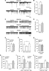
Alcohol dependence disrupted the role of extracellular calcium influx in acute alcohol-induced CeA activity. A, Representative sIPSC traces from CeA neurons of naive and CIE rats in baseline conditions and during acute alcohol (44 mm EtOH) superfusion. B, CIE rat CeA neurons (52 cells from 23 rats) have a higher baseline sIPSC frequency compared with naive rat cells (78 cells from 44 rats). C, Representative firing traces from CeA neurons of naive and CIE rats. D, The baseline firing rate was similar in cells from the CeA of naive (24 cells from 14 rats) and CIE rats (24 cells from 12 rats). E, EtOH significantly increased sIPSC frequencies in naive (18 cells from 13 rats) and CIE CeA neurons (7 cells from 4 rats). F, Cells treated twice with EtOH, with an intermediate 15 min wash period, showed no difference in EtOH enhancement of sIPSC frequency between the two applications (6 cells from 6 rats). G, EtOH significantly increased sIPSC frequencies in naive (8 cells from 5 rats) and CIE CeA neurons (8 cells from 4 rats) that were preloaded with 10 mm BAPTA. H, In naive rats, EtOH significantly increased the firing rate in 16/24 cells and decreased it in 8/24 cells (from 11 rats), whereas in CIE rats, EtOH increased the firing rate in 14/14 cells from 8 rats. I, EtOH significantly increased the sIPSC frequencies in naive and CIE CeA neurons exposed to 5 mm Ca2+ aCSF, and in CIE neurons exposed to 0.5 mm Ca2+ aCSF (normalized to pre-EtOH baseline). For these extracellular calcium experiments, 5–8 cells from a minimum of 4 rats were used for each experimental group. J, The sIPSC frequency was increased only in naive rat CeA neurons bathed in 5 mm Ca2+ aCSF (which is normalized to the 2 mm Ca2+ aCSF baseline). Five to 13 cells from a minimum of 4 rats were used for each experimental group. All data are normalized to a pre-EtOH baseline (or 2 mm Ca2+ aCSF baseline as specified) and presented as mean ± SEM. *p < 0.05, **p < 0.01, ***p < 0.001; ###p < 0.001. Ns, not significant.
Figure 2.
Figure 2.
LTCC mechanisms of alcohol-induced CeA activity are lost in CIE rats. A, Nifedipine (10 μm) prevented acute alcohol's (44 mm EtOH) enhancement of sIPSC frequency in naive rats (11 cells from 7 rats), but not in CIE rats (9 cells from 6 rats). B, The EtOH enhancement of neuronal firing was also blocked by nifedipine in naive rats (9 cells from 7 rats), but not in CIE rats (7 cells from 4 rats). C, D, In naive rats, previously EtOH-responsive cells no longer displayed EtOH enhancement of (C) sIPSC frequency (7 cells from 4 rats) or (D) neuronal firing (9 cells from 7 rats) in the presence of nifedipine (nif). For these multistage experiments, all groups are normalized to the original pre-EtOH baseline. E, EtOH's enhancement of sIPSC frequency was blocked in the presence of a second LTCC blocker, verapamil (100 μm verap; 6 cells from 4 rats), but was unchanged by the N-type voltage-gated calcium channel blocker ω-conotoxin GVIA (1 μm cono; 7 cells from 4 rats) or the P/Q-type voltage-gated calcium channel blocker, ω-agatoxin TK (500 nm aga; 4 cells from 3 rats). F, Top, Representative Western blot images of LTCC-subtype Cav1.2 membrane abundance from the CeA and mPFC of naive and CIE rats. Bottom, Quantification revealed a significant decrease in CeA Cav1.2 membrane expression in CIE versus naive rats (6 rats were used for each experimental group). G, R121919 (1 μm) prevented EtOH enhancement of sIPSC frequency in CIE rats (6 cells from 4 rats), but not in naive rats (9 cells from 4 rats). H, The EtOH enhancement of neuronal firing was also blocked by R121919 in CIE rats (5 cells from 4 rats), but not in naive rats (7 cells from 4 rats). I, IP3R blockade (42 μm 2-APB) prevented EtOH's enhancement of the sIPSC frequency in naive rats, but not in CIE rats, whereas RyR blockade (20 μm) produced the opposite result as it prevented EtOH's effects in CIE rats, but not naive rats. For these intracellular calcium experiments, 4–7 cells from a minimum of 3 rats were used for each experimental group. J, The PKA (10 μm Rp-cAMP) and PKC antagonists (200 nm Ro 32–0432) prevented EtOH enhancement of sIPSC frequencies in CIE rats, but not naive rats. For these protein kinase experiments, 5–6 cells from a minimum of 3 rats were used for each experimental group. All data are normalized to a pre-EtOH baseline and presented as mean ± SEM. *p < 0.05, **p < 0.01, ***p < 0.001.
Figure 3.
Figure 3.
Intra-CeA nifedipine reduces alcohol self-administration in nondependent rats. A, Effect of intra-CeA nifedipine (Nif) and R121919 (R12) on alcohol self-administration in nondependent rats (n = 8). Data represent the number of reinforced responses for alcohol (top) or water (bottom). B, Effect of intra-CeA Nif and R12 on alcohol self-administration in alcohol-dependent rats (n = 11) after their escalation of alcohol intake (Esc). C, Histological reconstruction showing correct (circles) and incorrect (triangles) injections into the CeA (drawing from the atlas of Paxinos and Watson, 2007). Three animals showed incorrect cannula placement and were excluded from the experiment. D–G, Effect of systemic Nif or R12 on alcohol self-administration in nondependent (n = 10) and alcohol-dependent rats (n = 12). Data presented as mean ± SEM. *p < 0.05 vs vehicle (Veh), **p < 0.01 vs Veh, ***p < 0.001 vs Veh, ###p < 0.001 vs baseline (Bsl).
Figure 4.
Figure 4.
Schematic illustrating how the LTTC-based mechanism that mediates the CeA's neuronal response to alcohol is altered with dependence, and the behavioral outcome of this neuroadaptation. Left, In naive/nondependent rats, an LTCC-based mechanism governs: (1) alcohol's enhancement of CeA action potential-dependent GABA release and (2) CeA-driven alcohol consumption. Right, In alcohol-dependent rats, these alcohol-induced effects on CeA activity and escalated alcohol intake are mediated by a CRF1-based mechanism.

References

    1. Alcami P, Franconville R, Llano I, Marty A (2012) Measuring the firing rate of high-resistance neurons with cell-attached recording. J Neuroscience 32:3118–3130. 10.1523/JNEUROSCI.5371-11.2012 - DOI - PMC - PubMed
    1. Altamura AC, Regazzetti MG, Porta M (1990) Nimodipine in human alcohol withdrawal syndrome–an open study. Eur Neuropsychopharmacol 1:37–40. 10.1016/0924-977X(90)90008-X - DOI - PubMed
    1. Bajo M, Cruz MT, Siggins GR, Messing R, Roberto M (2008) Protein kinase C epsilon mediation of CRF- and ethanol-induced GABA release in central amygdala. Proc Natl Acad Sci U S A 105:8410–8415. 10.1073/pnas.0802302105 - DOI - PMC - PubMed
    1. Banger M, Benkert O, Röschke J, Herth T, Hebenstreit M, Philipp M, Aldenhoff JB (1992) Nimodipine in acute alcohol withdrawal state. J Psychiatr Res 26:117–123. 10.1016/0022-3956(92)90003-7 - DOI - PubMed
    1. Bhat S, Dao DT, Terrillion CE, Arad M, Smith RJ, Soldatov NM, Gould TD (2012) CACNA1C (Cav1.2) in the pathophysiology of psychiatric disease. Prog Neurobiol 99:1–14. 10.1016/j.pneurobio.2012.06.001 - DOI - PMC - PubMed

MeSH terms

Substances