. 2017 Jun 1;10(6):787-796.
doi: 10.1242/dmm.025692. Epub 2017 Mar 31.

Progesterone induces neuroprotection following reperfusion-promoted mitochondrial dysfunction after focal cerebral ischemia in rats

Affiliations

Progesterone induces neuroprotection following reperfusion-promoted mitochondrial dysfunction after focal cerebral ischemia in rats

Syed Suhail Andrabi et al. Dis Model Mech. .

Abstract

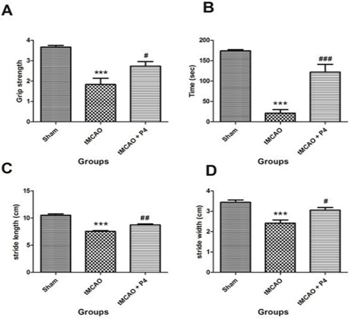
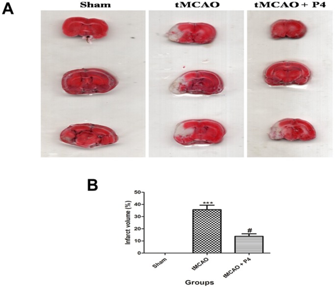
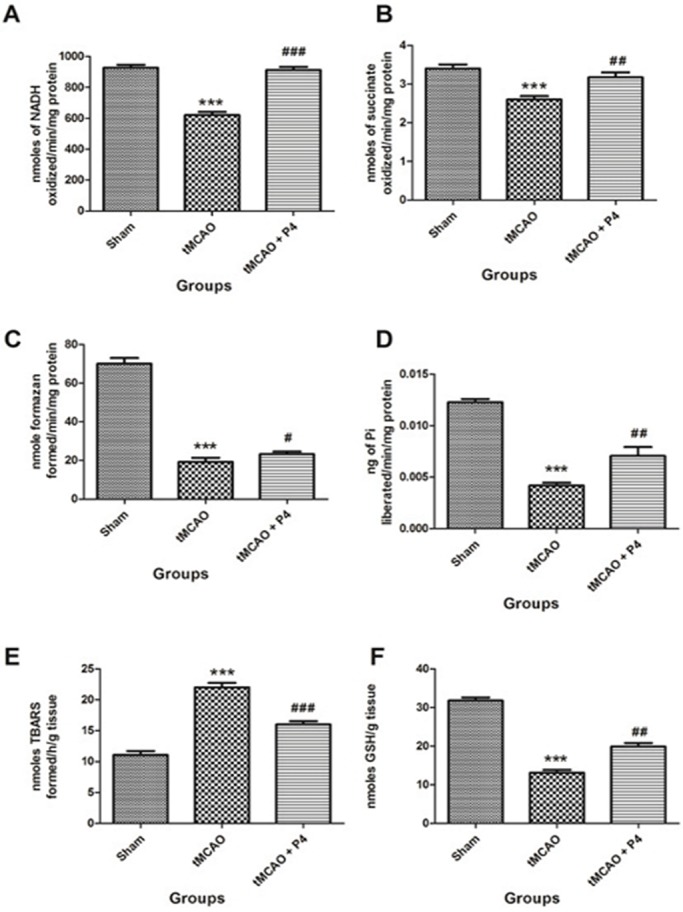
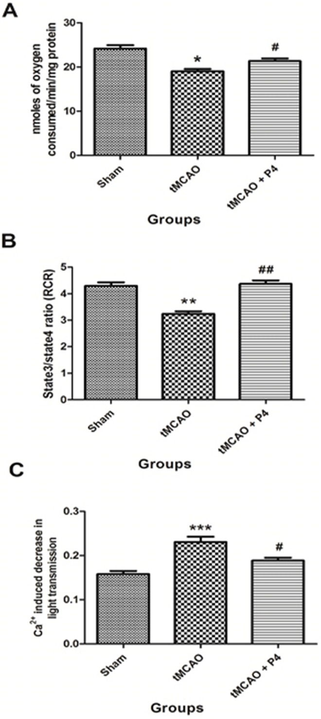
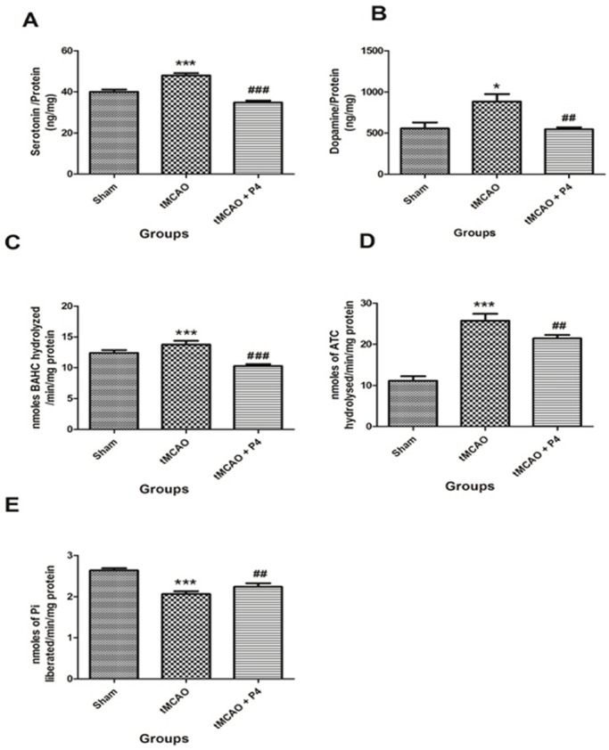
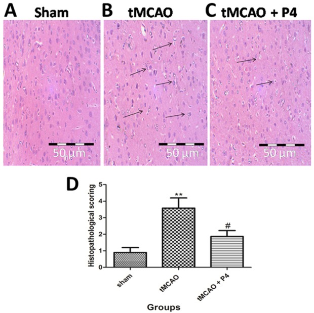
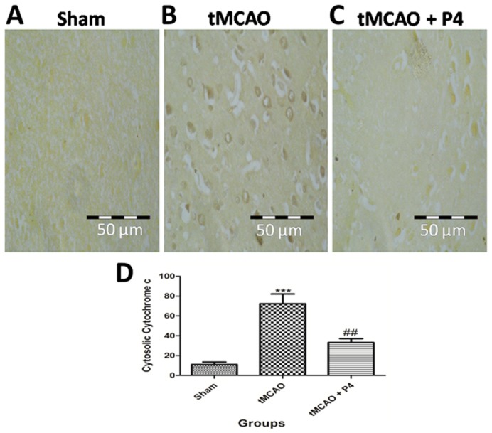
Organelle damage and increases in mitochondrial permeabilization are key events in the development of cerebral ischemic tissue injury because they cause both modifications in ATP turnover and cellular apoptosis/necrosis. Early restoration of blood flow and improvement of mitochondrial function might reverse the situation and help in recovery following an onset of stroke. Mitochondria and related bioenergetic processes can be effectively used as pharmacological targets. Progesterone (P4), one of the promising neurosteroids, has been found to be neuroprotective in various models of neurological diseases, through a number of mechanisms. This influenced us to investigate the possible role of P4 in the mitochondria-mediated neuroprotective mechanism in an ischemic stroke model of rat. In this study, we have shown the positive effect of P4 administration on behavioral deficits and mitochondrial health in an ischemic stroke injury model of transient middle cerebral artery occlusion (tMCAO). After induction of tMCAO, the rats received an initial intraperitoneal injection of P4 (8 mg/kg body weight) or vehicle at 1 h post-occlusion followed by subcutaneous injections at 6, 12 and 18 h. Behavioral assessment for functional deficits included grip strength, motor coordination and gait analysis. Findings revealed a significant improvement with P4 treatment in tMCAO animals. Staining of isolated brain slices from P4-treated rats with 2,3,5-triphenyltetrazolium chloride (TTC) showed a reduction in the infarct area in comparison to the vehicle group, indicating the presence of an increased number of viable mitochondria. P4 treatment was also able to attenuate mitochondrial reactive oxygen species (ROS) production, as well as block the mitochondrial permeability transition pore (mPTP), in the tMCAO injury model. In addition, it was also able to ameliorate the altered mitochondrial membrane potential and respiration ratio in the ischemic animals, thereby suggesting that P4 has a positive effect on mitochondrial bioenergetics. In conclusion, these results demonstrate that P4 treatment is beneficial in preserving the mitochondrial functions that are altered in cerebral ischemic injury and thus can help in defining better therapies.

Keywords: Apoptosis; Cerebral ischemia; Mitochondria; Neurobehavior; Neuroprotection; Progesterone.


Conflict of interest statement

Competing interestsThe authors declare no competing or financial interests.

Figures

Fig. 1.
Fig. 1.
Behavioral test parameters. All animals were subjected to various neurological tests. (A) Grip strength test done to assess the muscular strength. ***P<0.001 versus sham, #P<0.05 versus tMCAO. (B) Time remaining on the rotarod. ***P<0.001 versus sham, ###P<0.001 versus tMCAO. (C) Stride length to assess the gait impairment. ***P<0.001 versus sham, ##P<0.01 versus tMCAO. (D) Stride width. ***P<0.001 versus sham, #P<0.05 versus tMCAO (n=6 in each group).
Fig. 2.
Fig. 2.
Effect of P4 on infarct volume. (A) Images of TTC staining. In the tMCAO group, there was severe infarction as compared to that of the P4 group. Infarcts are shown as white (unstained) regions involving cortex. (B) Infarct volume. ***P<0.001 versus sham, #P<0.05 versus tMCAO.
Fig. 3.
Fig. 3.
Effect of P4 on mitochondrial complexes and oxidative parameters. Analysis of the levels of mitochondrial complexes in isolated mitochondria from the frontal cortex of the brain (n=6 in each group). (A) Effect of P4 on NADH dehydrogenase (complex I). ***P<0.001 versus sham, ###P<0.001 versus tMCAO. (B) Effect of progesterone on succinate dehydrogenase (complex II). ***P<0.001 versus sham, ##P<0.01 versus tMCAO. (C) MTT assay was done to assess the metabolic activity of cells in the frontal cortex of the brain (n=6). ***P<0.001 versus sham, #P<0.05 versus tMCAO. (D) Effect of P4 on F1-F0 synthase activity (complex V). ***P<0.001 versus sham, ##P<0.01 versus tMCAO. (E) Effect of P4 on mitochondrial lipid peroxidation. ***P<0.001 versus sham, ###P<0.001 versus tMCAO. (F) Effect of P4 on mitochondrial GSH. ***P<0.001 versus sham, ##P<0.01 versus tMCAO.
Fig. 4.
Fig. 4.
Effect of P4 on mitochondrial bioenergetics. (A) Effect of P4 on mitochondrial oxygen consumption (state 3 respiration rate). *P<0.05 versus sham, #P<0.05 versus tMCAO. (B) Effect of P4 on respiratory control ratio (RCR) in sham, tMCAO and tMCAO+P4 (n=6 in each group). **P<0.01 versus sham, ##P<0.01 versus tMCAO. (C) Effect of P4 on mitochondrial swelling. ***P<0.001 versus sham, #P<0.05 versus tMCAO (n=6 in each group).
Fig. 5.
Fig. 5.
Measurement of mitochondrial ROS and mitochondrial membrane potential (MMP). (A) In the FSC/SSC plot of the isolated mitochondria, 10,000 events were collected within gate R1. (B) Measurement of ROS production in sham, tMCAO and tMCAO+P4 as shown by changes in DCF fluorescence. (C) The relative changes in DCF fluorescence intensity. ***P<0.001 versus sham, #P<0.05 versus tMCAO. (D) Changes in MMP in sham, tMCAO and tMCAO+P4 as reflected by changes in TMRE fluorescence. (E) The relative changes in TMRE fluorescence intensity are shown. **P<0.001 versus sham, #P<0.05 versus tMCAO. n=6 in each group.
Fig. 6.
Fig. 6.
Effect of P4 on 5-HT, dopamine and neurotoxicity parameters. (A) Effect of P4 on 5-HT. ***P<0.001 versus sham, ###P<0.001 versus tMCAO. (B) Effect of P4 on dopamine. *P<0.05 versus sham, ##P<0.01 versus tMCAO (n= 6 in each group). (C) Effect of P4 on monoamine oxidase activity. ***P<0.001 versus sham, ###P<0.001 versus tMCAO. (D) Effect of P4 on acetylcholine esterase activity. ***P<0.001 versus sham, ##P<0.01 versus tMCAO. (E) Effect of P4 on Na+ K+-ATPase. ***P<0.001 versus sham, ##P<0.01 versus tMCAO (n=6 in each group).
Fig. 7.
Fig. 7.
Effect of P4 on histopathology. (A-C) Representative histopathological photomicrograph of frontoparietal layers of the sham, tMCAO and tMCAO+P4 group. In the sham group, there was no vacuolation or any neuronal loss. In the tMCAO group, there was vacuolation and heavy neuronal loss. In the P4 treated group there was the partial neuronal loss. Magnification at 40×. (D) Histological alterations are represented graphically, showing significant changes (**P<0.001) in tMCAO and that, in the P4 treated group, there was a significant improvement (#P<0.05).
Fig. 8.
Fig. 8.
Effect of P4 on cytochrome c translocation. (A-C) Representative images of the frontoparietal layers of the brain were taken for analysis of the translocation of cytochrome c from mitochondria to cytosol. In the tMCAO group, the cytochrome c immunostaining is higher as compared to that in the sham group, which suggests cytosolic translocation of cytochrome c. Treatment with P4 is able to reduce the translocation of cytochrome c following tMCAO. (D) Quantitative measurements of cytosolic cytochrome c release. Cytosolic translocation of cytochrome c was found to be significant (***P<0.001) in tMCAO rats and its translocation was significantly inhibited (##P<0.01) with P4 treatment.

References

    1. Affonso A. C., Machado D. G., Malgarin F., Fraga D. B., Ghedim F., Zugno A., Streck E. L., Schuck P. F. and Ferreira G. C. (2013). Increased susceptibility of brain acetylcholinesterase activity to methylmalonate in young rats with renal failure. Metab. Brain Dis. 28, 493-500. 10.1007/s11011-013-9396-0 - DOI - PubMed
    1. Aggarwal R., Medhi B., Pathak A., Dhawan V. and Chakrabarti A. (2008). Neuroprotective effect of progesterone on acute phase changes induced by partial global cerebral ischaemia in mice. J. Pharm. Pharmacol. 60, 731-737. 10.1211/jpp.60.6.0008 - DOI - PubMed
    1. Andrabi S. S., Parvez S. and Tabassum H. (2015). Melatonin and ischemic stroke: mechanistic roles and action. Adv. Pharmacol. Sci. 2015, 384750 10.1155/2015/384750 - DOI - PMC - PubMed
    1. Ashafaq M., Khan M. M., Shadab Raza S., Ahmad A., Khuwaja G., Javed H., Khan A., Islam F., Siddiqui M. S., Safhi M. M. et al. (2012a). S-allyl cysteine mitigates oxidative damage and improves neurologic deficit in a rat model of focal cerebral ischemia. Nutr. Res. 32, 133-143. 10.1016/j.nutres.2011.12.014 - DOI - PubMed
    1. Ashafaq M., Raza S. S., Khan M. M., Ahmad A., Javed H., Ahmad M. E., Tabassum R., Islam F., Siddiqui M. S., Safhi M. M. et al. (2012b). Catechin hydrate ameliorates redox imbalance and limits inflammatory response in focal cerebral ischemia. Neurochem. Res. 37, 1747-1760. 10.1007/s11064-012-0786-1 - DOI - PubMed

Publication types

MeSH terms