. 2017 May 15;130(10):1752-1759.
doi: 10.1242/jcs.198358. Epub 2017 Mar 31.

Spatial distribution and molecular dynamics of dystrophin glycoprotein components at the neuromuscular junction in vivo

Affiliations

Spatial distribution and molecular dynamics of dystrophin glycoprotein components at the neuromuscular junction in vivo

Mohamed Aittaleb et al. J Cell Sci. .

Abstract

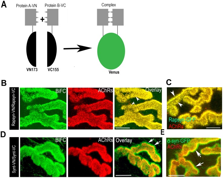
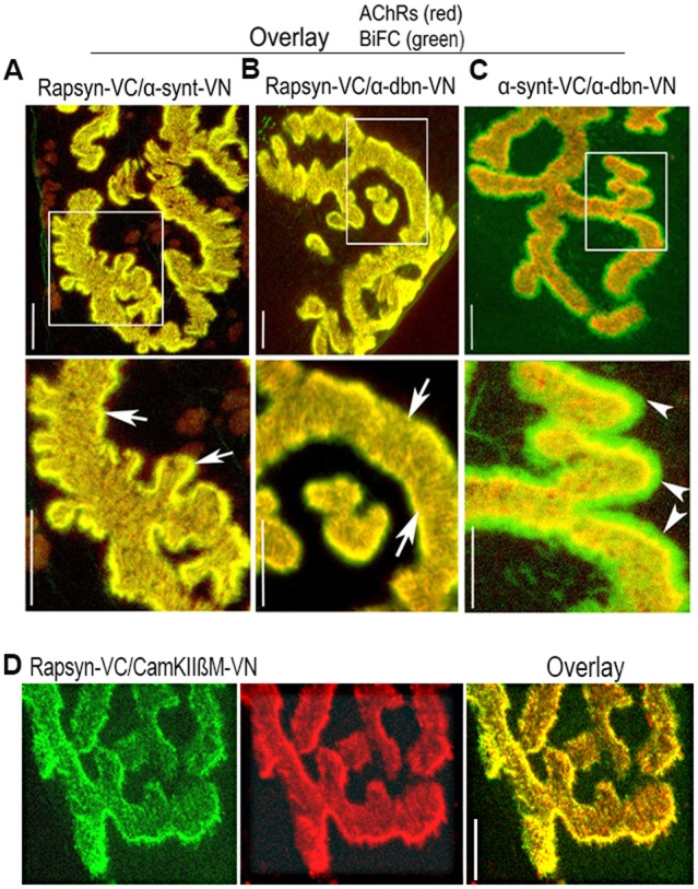
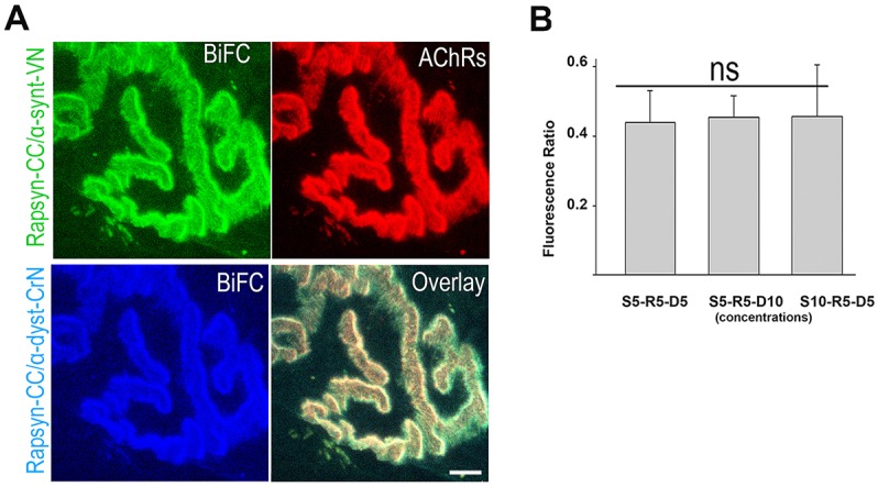
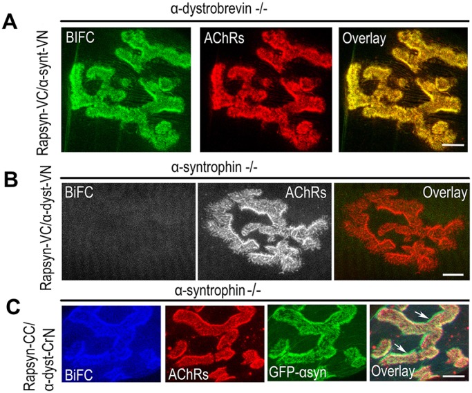
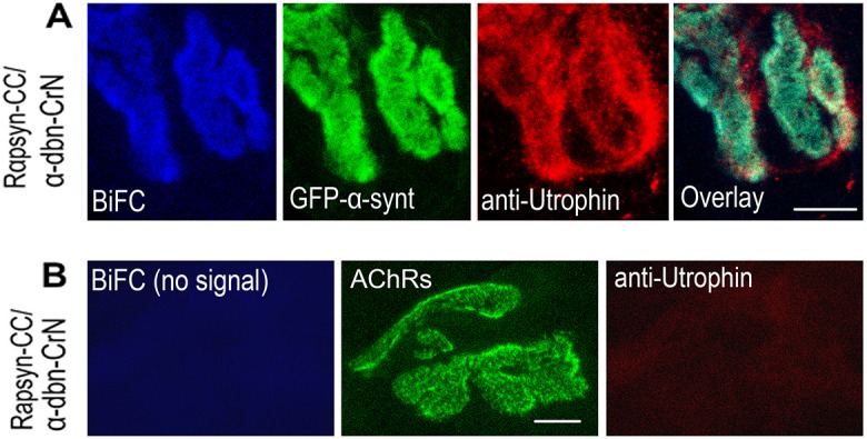
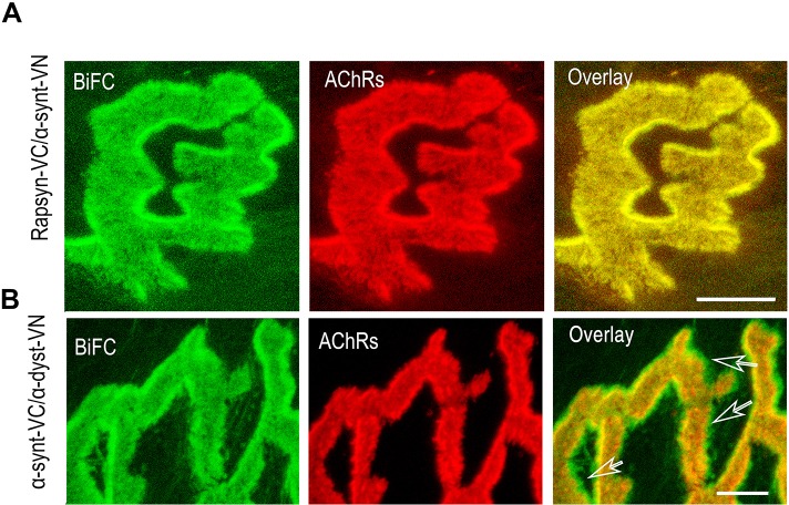
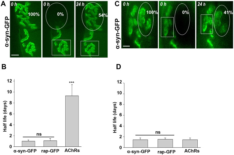
A bimolecular fluorescence complementation (BiFC) approach was used to study the molecular interactions between different components of the postsynaptic protein complex at the neuromuscular junction of living mice. We show that rapsyn forms complex with both α-dystrobrevin and α-syntrophin at the crests of junctional folds. The linkage of rapsyn to α-syntrophin and/or α-dystrobrevin is mediated by utrophin, a protein localized at acetylcholine receptor (AChR)-rich domains. In mice deficient in α-syntrophin, in which utrophin is no longer present at the synapse, rapsyn interaction with α-dystrobrevin was completely abolished. This interaction was completely restored when either utrophin or α-syntrophin was introduced into muscles deficient in α-syntrophin. However, in neuromuscular junctions deficient in α-dystrobrevin, in which utrophin is retained, complex formation between rapsyn and α-syntrophin was unaffected. Using fluorescence recovery after photobleaching, we found that α-syntrophin turnover is 5-7 times faster than that of AChRs, and loss of α-dystrobrevin has no effect on rapsyn and α-syntrophin half-life, whereas the half-life of AChR was significantly altered. Altogether, these results provide new insights into the spatial distribution of dystrophin glycoprotein components and their dynamics in living mice.

Keywords: AChR; DGC; Dystrophin glycoprotein complex; NMJ; Neuromuscular junction.


Conflict of interest statement

Competing interestsThe authors declare no competing or financial interests.

Figures

Fig. 1.
Fig. 1.
In vivo BiFC shows self-interaction between rapsyn–VN and rapsyn–VC, and between α-syntrophin–VN and α-syntrophin–VC at the NMJ. (A) Schematic representation of BiFC assay. Association of two non-fluorescent protein fragments that are fused to proteins; when they come close to each other, the native three-dimensional structure is formed and emits a fluorescence signal. (B–E) Sternomastoid muscles were electroporated with constructs expressing either rapsyn–VN and rapsyn–VC, α-syntrophin–VC (Synt–VC) and α-syntrophin–VN (Synt–VN), rapsyn–GFP, or α-syntrophin–GFP, and NMJs were identified by labeling AChRs with BTX–Alexa594 (red). (B) A representative image showing BiFC signal of rapsyn–VC and rapsyn–VN. (C) A representative image of an NMJ expressing rapsyn–GFP fusion protein. Note that BiFC and rapsyn–GFP signals localized perfectly with AChRs labeled with BTX–Alexa594 at synaptic sites (see arrowheads). (D) A representative image showing the BiFC signal of α-syntrophin–VC with α-syntrophin–VN. (E) A representative image of an NMJ expressing α-syntrophin–GFP. Note that BiFC and GFP signals are present in crests and at the bottom of the junctional folds (see arrows). Six mice were used in each experiment. Scale bars: 5 µm.
Fig. 2.
Fig. 2.
In vivo BiFC shows interaction between rapsyn, α-dystrobrevin, α-syntrophin and CamKIIβM at the NMJ. Sternomastoid muscles were co-electroporated either with rapsyn–VC and α-syntrophin–VN (α-synt–VN), or rapsyn–VC and α-dystrobrevin–VN (α-dbn–VN), or α-dystrobrevin–VN and α-syntrophin–VC (α-synt–VC); 7 days later, muscles were bathed with BTX–Alexa594 to label AChRs, and synapses expressing BiFC signals were imaged. (A) A representative synapse showing that the fluorescence complementation between rapsyn–VC and α-syntrophin–VN is restricted to the crests of junctional folds where they precisely colocalized with AChRs (see arrows). (B) A representative synapse image showing fluorescence complementation between rapsyn–VC and α-dystrobrevin–VN (α-dbn–VN) only at the crests of junctional folds that were identified by AChR labeling (see arrows). (C) A representative synapse image showing the fluorescence complementation between α-syntrophin and α-dystrobrevin at both the crest and troughs of junctional folds (see arrowheads). Boxed areas (above) are enlarged in the images below. (D) A representative image showing the BiFC signal between rapsyn–VC and CamKIIβM–VN. Scale bars: 5 µm. Eight mice were used in each experiment.
Fig. 3.
Fig. 3.
Rapsyn interacts with both α-syntrophin and α-dystrobrevin. The sternomastoid muscle was co-electroporated with a fixed amount of rapsyn–CC with one of the following: (i) the same amount of α-syntrophin–VN and α-dystrobrevin–CrN (α-dyst–CrN) (S5-R5-D5); (ii) a low concentration of α-syntrophin–VN (α-synt–VN) and high concentration of α-dystrobrevin–CrN (S5-R5-D10); (iii) or a high concentration of α-syntrophin–VN and low concentration of α-dystrobrevin–CrN (S10-R5-D5). After 7 days, the muscle was bathed with BTX–Alexa594 to label AChRs, fixed and imaged. (A) A representative image of a synapse showing the complementation of rapsyn–CC with α-syntrophin–VN, and rapsyn–CC with α-dystrobrevin–CrN (α-dbn-CrN). (B) Graph showing quantification of BiFC signals between rapsyn–CC and α-syntrophin–VN and rapsyn–CC and α-dystrobrevin–CrN at different concentrations. Note that the fluorescence ratio value between green and cyan remains the same between different constructs concentrations. Each bar represents the mean±s.d. of 40 junctions (six mice). Scale bar: 5 µm. One-way ANOVA test, P=0.89; ns, not significant. R, rapsyn; S, α-syntrophin; D, α-dystrobrevin; numbers indicate the amount of plasmid (µg).
Fig. 4.
Fig. 4.
In vivo BiFC complementation between rapsyn and α-dystrobrevin is abolished at NMJs of mice deficient in α-syntrophin but not in synapses deficient in α-dystrobrevin. (A) The sternomastoid muscle of mice deficient in α-dystrobrevin was electroporated with rapsyn–VC and α-syntrophin–VN constructs (α-synt–VN); 7 days later, AChRs on muscles were labeled with BTX–Alexa594, fixed and imaged. A representative synapse expressing the BiFC signal is shown. Note that the BiFC signal at the NMJ is not impaired by the absence of α-dystrobrevin. (B) A representative synapse from the sternomastoid muscle of mice deficient in α-syntrophin that had been co-electroporated with rapsyn–VC and α-dystrobrevin–VN (α-dyst–VN) constructs were bathed with BTX–Alexa594 to label AChRs. Note the complete absence of BiFC signal at the NMJ. (C) A representative synapse from the sternomastoid muscle of mice deficient in α-syntrophin that had been electroporated with rapsyn–CC, α-dystrobrevin–CrN (α-dyst–CrN) and α-syntrophin–GFP (GFP–αsyn). Note that the BiFC signal at the synapses was rescued at NMJs expressing α-syntrophin–GFP. AChRs were labeled with BTX–Alexa594. Scale bars: 5 µm. Seven mice were used in each experiment.
Fig. 5.
Fig. 5.
Utrophin expression is required for the fluorescence complementation between rapsyn and α-syntrophin and α-dystrobrevin. The sternomastoid muscle of mice deficient in α-syntrophin was co-electroporated with rapsyn–CC, α-dystrobrevin–CrN (α-dbn–CrN), α-syntrophin–GFP (GFP–α-synt) and utrophin. (A) Examples of high-resolution confocal images of an NMJ expressing α-syntrophin–GFP and BiFC signal between rapsyn–CC and α-dystrobrevin–CrN are shown. (B) Examples of an NMJ imaged from the sternomastoid muscle of mice deficient in α-syntrophin that had been co-electroporated with rapsyn–CC and α-dystrobrevin–CrN, and 7 days after electroporation, muscles were fixed and stained with antibody against utrophin and BTX–Alexa488. Note that there is no fluorescence complementation between rapsyn and α-dystrobrevin and no utrophin expression in the absence of α-syntrophin. Scale bars: 5 µm. Eight mice were used in each experiment.
Fig. 6.
Fig. 6.
Postsynaptic rapsyn, α-syntrophin and α-dystrobrevin are targeted to their specific locations as individual proteins. The sternomastoid muscle was electroporated with either rapsyn–VC and α-syntrophin–VN (Rapsyn–VC/α-synt-VN) or with α-syntrophin–VC and α-dystrobrevin–VN (α–synt–VC/α–dyst-VN). The muscle was bathed with BTX–Alexa594 to label AChRs, then fixed and imaged. (A) A representative image of a synapse showing fluorescence complementation between rapsyn and α-syntrophin at the crest of synaptic folds only, as indicated by the perfect colocalization of BiFC signal with AChR at the crest of synaptic folds of the synapses. (B) Example of a high-resolution confocal image of an NMJ expressing the BiFC signal of α-dystrobrevin and α-syntrophin at the crests and bottom of the junctional folds (see arrows). Scale bars: 5 µm. Six mice were used in each experiment.
Fig. 7.
Fig. 7.
The DGC α-syntrophin half-life at wild-type and α-dystrobrevin mutant NMJs. Sternomastoid muscles of live wild-type and α-dystrobrevin mutant mice that had been electroporated with either rapsyn–GFP (rap–GFP) or α-syntrophin–GFP (α-syn–GFP); 7 days later, the fluorescence from discrete regions of NMJs expressing GFP was removed with a laser and re-imaged. The recovery of fluorescence was monitored over 24 h. Notice that only images of sternomastoid muscles electroporated with α-syntrophin–GFP are shown. (A) Example of a wild-type NMJ that was electroporated with α-syntrophin–GFP, imaged at time 0 and immediately bleached and re-imaged at 24 h (eight mice). (B) Graph summarizing data of the half-time of α-syntrophin, rapsyn and AChRs obtained from 47 synapses. (C) Example of NMJ deficient in α-dystrobrevin expressing α-syntrophin–GFP that was imaged at time 0 and then immediately bleached and re-imaged at 24 h. (D) Graph summarizing data of half-time of α-syntrophin, rapsyn and AChRs obtained from 42 mutant synapses (12 mice). All data represent mean±s.d. Scale bars: 10 µm.

Similar articles

Cited by

References

    1. Adams M. E., Kramarcy N., Krall S. P., Rossi S. G., Rotundo R. L., Sealock R. and Froehner S. C. (2000). Absence of alpha-syntrophin leads to structurally aberrant neuromuscular synapses deficient in utrophin. J. Cell Biol. 150, 1385-1398. 10.1083/jcb.150.6.1385 - DOI - PMC - PubMed
    1. Adams M. E., Anderson K. N. E. and Froehner S. C. (2010). The alpha-syntrophin PH and PDZ domains scaffold acetylcholine receptors, utrophin, and neuronal nitric oxide synthase at the neuromuscular junction. J. Neurosci. 30, 11004-11010. 10.1523/JNEUROSCI.1930-10.2010 - DOI - PMC - PubMed
    1. Ahn A. H., Freener C. A., Gussoni E., Yoshida M., Ozawa E. and Kunkel L. M. (1996). The three human syntrophin genes are expressed in diverse tissues, have distinct chromosomal locations, and each bind to dystrophin and its relatives. J. Biol. Chem. 271, 2724-2730. 10.1074/jbc.271.5.2724 - DOI - PubMed
    1. Akaaboune M., Culican S. M., Turney S. G. and Lichtman J. W. (1999). Rapid and reversible effects of activity on acetylcholine receptor density at the neuromuscular junction in vivo. Science 286, 503-507. 10.1126/science.286.5439.503 - DOI - PubMed
    1. Akaaboune M., Grady R. M., Turney S., Sanes J. R. and Lichtman J. W. (2002). Neurotransmitter receptor dynamics studied in vivo by reversible photo-unbinding of fluorescent ligands. Neuron 34, 865-876. 10.1016/S0896-6273(02)00739-0 - DOI - PubMed

Publication types