

Cardiac Fibroblast Activation Post-Myocardial Infarction: Current Knowledge Gaps
- PMID: 28365093
- PMCID: PMC5437868
- DOI: 10.1016/j.tips.2017.03.001
Cardiac Fibroblast Activation Post-Myocardial Infarction: Current Knowledge Gaps
Abstract
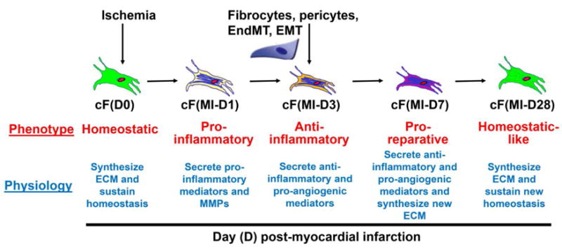
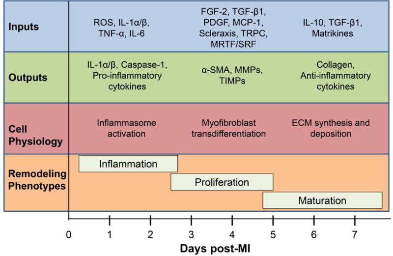
In response to myocardial infarction (MI), the wound healing response of the left ventricle (LV) comprises overlapping inflammatory, proliferative, and maturation phases, and the cardiac fibroblast is a key cell type involved in each phase. It has recently been appreciated that, early post-MI, fibroblasts transform to a proinflammatory phenotype and secrete cytokines and chemokines as well as matrix metalloproteinases (MMPs). Later post-MI, fibroblasts are activated to anti-inflammatory and proreparative phenotypes and generate anti-inflammatory and proangiogenic factors and extracellular matrix (ECM) components that form the infarct scar. Additional studies are needed to systematically examine how fibroblast activation shifts over the timeframe of the MI response and how modulation at different activation stages could alter wound healing and LV remodeling in distinct ways. This review summarizes current fibroblast knowledge as the foundation for a discussion of existing knowledge gaps.
Keywords: big data; computational models; extracellular matrix; fibroblast; inflammation; myocardial infarction; omics.
Copyright © 2017 Elsevier Ltd. All rights reserved.
Conflict of interest statement
None.
Figures
References
-
- Writing Group, M. et al. Heart Disease and Stroke Statistics-2016 Update: A Report From the American Heart Association. Circulation. 2016;133:e38–360. - PubMed
-
- Mikawa T, Gourdie RG. Pericardial mesoderm generates a population of coronary smooth muscle cells migrating into the heart along with ingrowth of the epicardial organ. Dev Biol. 1996;174:221–232. - PubMed
-
- Ali SR, et al. Developmental heterogeneity of cardiac fibroblasts does not predict pathological proliferation and activation. Circ Res. 2014;115:625–635. - PubMed
Publication types
MeSH terms
Grants and funding
LinkOut - more resources
Full Text Sources
Other Literature Sources
Medical
