

Benign Yellow Dot Maculopathy: A New Macular Phenotype
- PMID: 28366503
- PMCID: PMC5503697
- DOI: 10.1016/j.ophtha.2017.02.026
Benign Yellow Dot Maculopathy: A New Macular Phenotype
Abstract
Purpose: To describe a novel macular phenotype that is associated with normal visual function.
Design: Retrospective, observational case series.
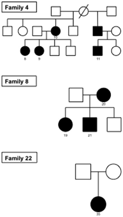
Participants: Thirty-six affected individuals from 23 unrelated families.
Methods: This was a retrospective study of patients who had a characteristic macular phenotype. Subjects underwent a full ocular examination, electrophysiologic studies, spectral-domain optical coherence tomography (OCT), and fundus autofluorescence imaging. Genomic analyses were performed using haplotype sharing analysis and whole-exome sequencing.
Main outcome measures: Visual acuity, retinal features, electroretinography, and whole-exome sequencing.
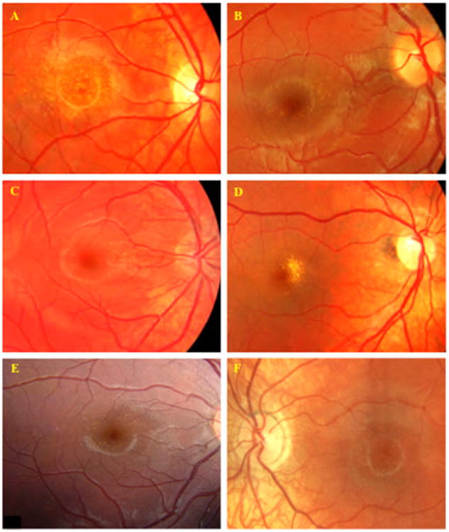
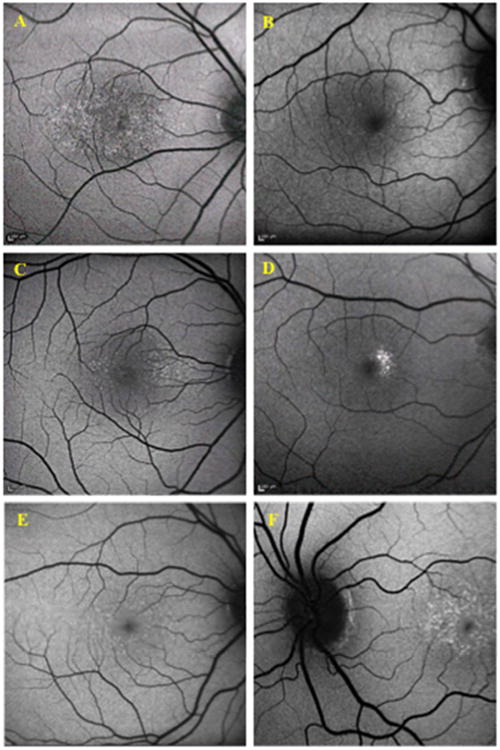
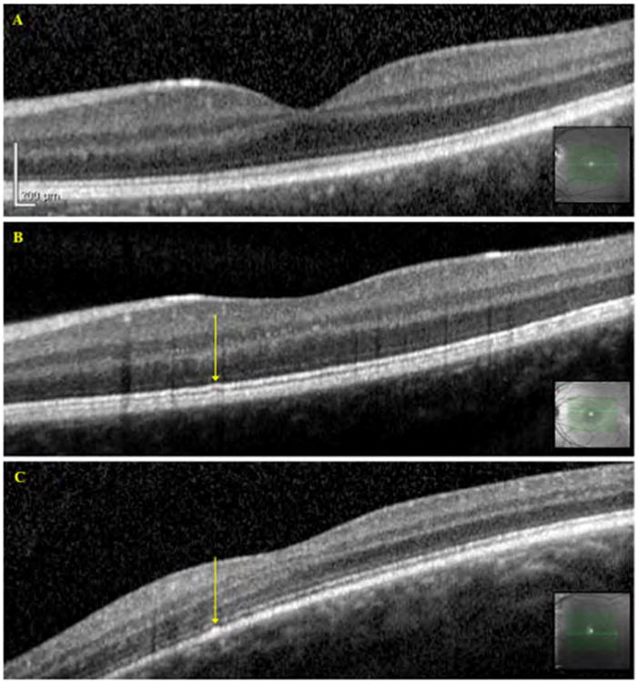
Results: Twenty-six of 36 subjects were female. The median age of subjects at presentation was 15 years (range, 5-59 years). The majority of subjects were asymptomatic and presented after a routine eye examination (22/36 subjects) or after screening because of a positive family history (13/36 subjects) or by another ophthalmologist (1/36 subjects). Of the 3 symptomatic subjects, 2 had reduced visual acuity secondary to nonorganic visual loss and bilateral ametropic amblyopia with strabismus. Visual acuity was 0.18 logarithm of the minimum angle of resolution (logMAR) or better in 30 of 33 subjects. Color vision was normal in all subjects tested, except for the subject with nonorganic visual loss. All subjects had bilateral symmetric multiple yellow dots at the macula. In the majority of subjects, these were evenly distributed throughout the fovea, but in 9 subjects they were concentrated in the nasal parafoveal area. The dots were hyperautofluorescent on fundus autofluorescence imaging. The OCT imaging was generally normal, but in 6 subjects subtle irregularities at the inner segment ellipsoid band were seen. Electrophysiologic studies identified normal macular function in 17 of 19 subjects and normal full-field retinal function in all subjects. Whole-exome analysis across 3 unrelated families found no pathogenic variants in known macular dystrophy genes. Haplotype sharing analysis in 1 family excluded linkage with the North Carolina macular dystrophy (MCDR1) locus.
Conclusions: A new retinal phenotype is described, which is characterized by bilateral multiple early-onset yellow dots at the macula. Visual function is normal, and the condition is nonprogressive. In familial cases, the phenotype seems to be inherited in an autosomal dominant manner, but a causative gene is yet to be ascertained.
Copyright © 2017 American Academy of Ophthalmology. Published by Elsevier Inc. All rights reserved.
Conflict of interest statement
Figures
References
-
- Moore AT. Childhood macular dystrophies. Current opinion in ophthalmology. 2009;20(5):363–8. - PubMed
-
- Michaelides M, Jeffery G, Moore AT. Developmental macular disorders: phenotypes and underlying molecular genetic basis. Br J Ophthalmol. 2012;96(7):917–24. - PubMed
-
- O'Sullivan J, et al. A paradigm shift in the delivery of services for diagnosis of inherited retinal disease. Journal of medical genetics. 2012;49(5):322–6. - PubMed
Publication types
MeSH terms
Substances
Grants and funding
LinkOut - more resources
Full Text Sources
Other Literature Sources
Medical
