Hepatoprotective effects of rice-derived peptides against acetaminophen-induced damage in mice
- PMID: 28366990
- PMCID: PMC5370527
- DOI: 10.3164/jcbn.16-44
Hepatoprotective effects of rice-derived peptides against acetaminophen-induced damage in mice
Abstract
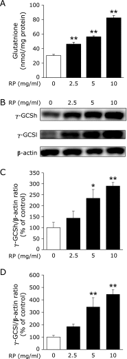
Glutathione, the most abundant intracellular antioxidant, protects cells against reactive oxygen species induced oxidative stress and regulates intracellular redox status. We found that rice peptides increased intracellular glutathione levels in human hepatoblastoma HepG2 cells. Acetaminophen is a commonly used analgesic. However, an overdose of acetaminophen causes severe hepatotoxicity via depletion of hepatic glutathione. Here, we investigated the protective effects of rice peptides on acetaminophen-induced hepatotoxicity in mice. ICR mice were orally administered rice peptides (0, 100 or 500 mg/kg) for seven days, followed by the induction of hepatotoxicity via intraperitoneal injection of acetaminophen (700 mg/kg). Pretreatment with rice peptides significantly prevented increases in serum alanine aminotransferase, aspartate aminotransferase, and lactate dehydrogenase levels and protected against hepatic glutathione depletion. The expression of γ-glutamylcysteine synthetase, a key regulatory enzyme in the synthesis of glutathione, was decreased by treatment with acetaminophen, albeit rice peptides treatment recovered its expression compared to that achieved treatment with acetaminophen. In addition, histopathological evaluation of the livers also revealed that rice peptides prevented acetaminophen-induced centrilobular necrosis. These results suggest that rice peptides increased intracellular glutathione levels and could protect against acetaminophen-induced hepatotoxicity in mice.
Keywords: acetaminophen; glutathione; liver injury; rice-derived peptide.
Conflict of interest statement
No potential conflicts of interest were disclosed.
Figures


References
-
- Meister A, Anderson ME. Glutathione. Annu Rev Biochem. 1983;52:711–760. - PubMed
-
- Reid M, Jahoor F. Glutathione in disease. Curr Opin Clin Nutr Metab Care. 2001;4:65–71. - PubMed
-
- Witschi A, Reddy S, Stofer B, Lauterburg BH. The systemic availability of oral glutathione. Eur J Clin Pharmacol. 1992;43:667–669. - PubMed
-
- Laskin DL, Pilaro AM. Potential role of activated macrophages in acetaminophen hepatotoxicity. I. Isolation and characterization of activated macrophages from rat liver. Toxicol Appl Pharmacol. 1986;86:204–215. - PubMed
-
- Hazai E, Vereczkey L, Monostory K. Reduction of toxic metabolite formation of acetaminophen. Biochem Biophys Res Commun. 2002;291:1089–1094. - PubMed
LinkOut - more resources
Full Text Sources
Other Literature Sources
Molecular Biology Databases
Research Materials

