Review
. 2017 Mar 17:11:103.
doi: 10.3389/fnhum.2017.00103. eCollection 2017.

Neurologic Correlates of Gait Abnormalities in Cerebral Palsy: Implications for Treatment

Affiliations
Review

Neurologic Correlates of Gait Abnormalities in Cerebral Palsy: Implications for Treatment

Joanne Zhou et al. Front Hum Neurosci. .

Abstract

Cerebral palsy (CP) is the most common movement disorder in children. A diagnosis of CP is often made based on abnormal muscle tone or posture, a delay in reaching motor milestones, or the presence of gait abnormalities in young children. Neuroimaging of high-risk neonates and of children diagnosed with CP have identified patterns of neurologic injury associated with CP, however, the neural underpinnings of common gait abnormalities remain largely uncharacterized. Here, we review the nature of the brain injury in CP, as well as the neuromuscular deficits and subsequent gait abnormalities common among children with CP. We first discuss brain injury in terms of mechanism, pattern, and time of injury during the prenatal, perinatal, or postnatal period in preterm and term-born children. Second, we outline neuromuscular deficits of CP with a focus on spastic CP, characterized by muscle weakness, shortened muscle-tendon unit, spasticity, and impaired selective motor control, on both a microscopic and functional level. Third, we examine the influence of neuromuscular deficits on gait abnormalities in CP, while considering emerging information on neural correlates of gait abnormalities and the implications for strategic treatment. This review of the neural basis of gait abnormalities in CP discusses what is known about links between the location and extent of brain injury and the type and severity of CP, in relation to the associated neuromuscular deficits, and subsequent gait abnormalities. Targeted treatment opportunities are identified that may improve functional outcomes for children with CP. By providing this context on the neural basis of gait abnormalities in CP, we hope to highlight areas of further research that can reduce the long-term, debilitating effects of CP.

Keywords: brain injury; cerebral palsy; gait; neuroimaging; neuromuscular deficits.


Figures
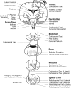
FIGURE 1
FIGURE 1
Brain regions and WM motor tracts affected in spastic, dyskinetic, and ataxic CP. A portion of the motor homunculus is superimposed on the cortex. Representations of the lateral ventricles, subcortical nuclei, cerebellum, brain stem, and rubrospinal and CSTs are outlined. The vermis of the cerebellum (posterior to the brainstem) is represented by dotted lines. Regions where the reticulospinal and vestibulospinal tracts descend through each layer of the brainstem and the spinal cord are shaded.
FIGURE 2
FIGURE 2
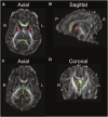
Diffusion-weighted image of an infant, highlighting brain regions of interest commonly affected in CP, using the semi automated JHU neonatal atlas (http://cmrm.med.jhmi.edu/cmrm/Data_neonate_atlas/atlas_neonate.htm). Axial view (A) and sagittal view (B) of the genu (light-green) and splenium (dark-green) of the corpus callosum, and anterior (red) and posterior (blue) limbs of the internal capsule. An axial view (C) and coronal view (D) highlight the thalamus (green) and globus pallidus (cyan). A, anterior; P, posterior; R, right; L, left. Figure reprinted with permission from Rose et al. (2015) Pediatric Research. http://www.nature.com/pr/journal/v78/n6/fig_tab/pr2015157f2.html
FIGURE 3
FIGURE 3
The gait cycle showing muscles that are influenced by neuromuscular deficits and contribute to gait abnormalities in spastic CP, in relation to phases of the gait cycle. IC, initial contact; OTO, opposite toe-off; OIC, opposite foot initial contact; TO, toe-off.

Similar articles

Cited by

References

    1. Ahlin K., Himmelmann K., Hagberg G., Kacerovsky M., Cobo T., Wennerholm U. B., et al. (2013). Cerebral palsy and perinatal infection in children born at term. Obstet. Gynecol. 122 41–49. 10.1097/AOG.0b013e318297f37f - DOI - PubMed
    1. Aravamuthan B. R., Waugh J. L. (2016). Localization of basal ganglia and thalamic damage in dyskinetic cerebral palsy. Pediatr. Neurol. 54 11–21. 10.1016/j.pediatrneurol.2015.10.005 - DOI - PubMed
    1. Arens L. J., Molteno C. D. (1989). A comparative study of postnatally-acquired cerebral palsy in cape town. Dev. Med. Child Neurol. 31 246–254. 10.1111/j.1469-8749.1989.tb03985.x - DOI - PubMed
    1. Arnold A. S., Liu M. Q., Schwartz M. H., Ounpuu S., Delp S. L. (2006). The role of estimating muscle-tendon lengths and velocities of the hamstrings in the evaluation and treatment of crouch gait. Gait Posture 23 273–281. 10.1016/j.gaitpost.2005.03.003 - DOI - PubMed
    1. Australian Cerebral Palsy Register Report [ACP] (2013). Report of the Australian Cerebral Palsy Register, Birth Years 1993–2006. Sydney: Cerebral Palsy Alliance Research Institute.

LinkOut - more resources