

Integrin β1 Increases Stem Cell Survival and Cardiac Function after Myocardial Infarction
- PMID: 28367125
- PMCID: PMC5355448
- DOI: 10.3389/fphar.2017.00135
Integrin β1 Increases Stem Cell Survival and Cardiac Function after Myocardial Infarction
Abstract
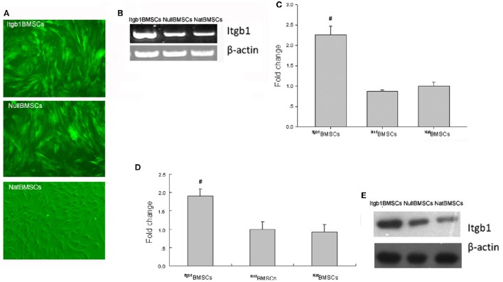
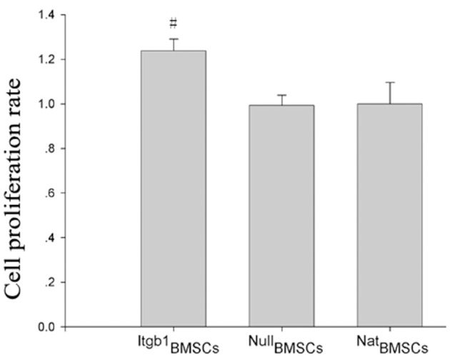
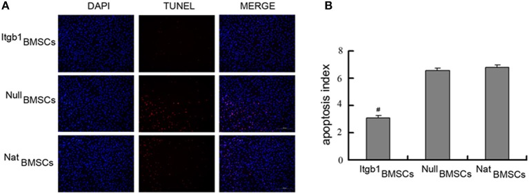
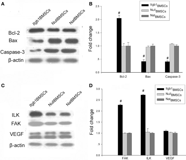
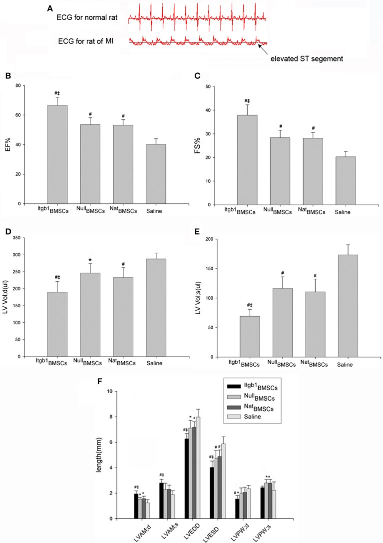
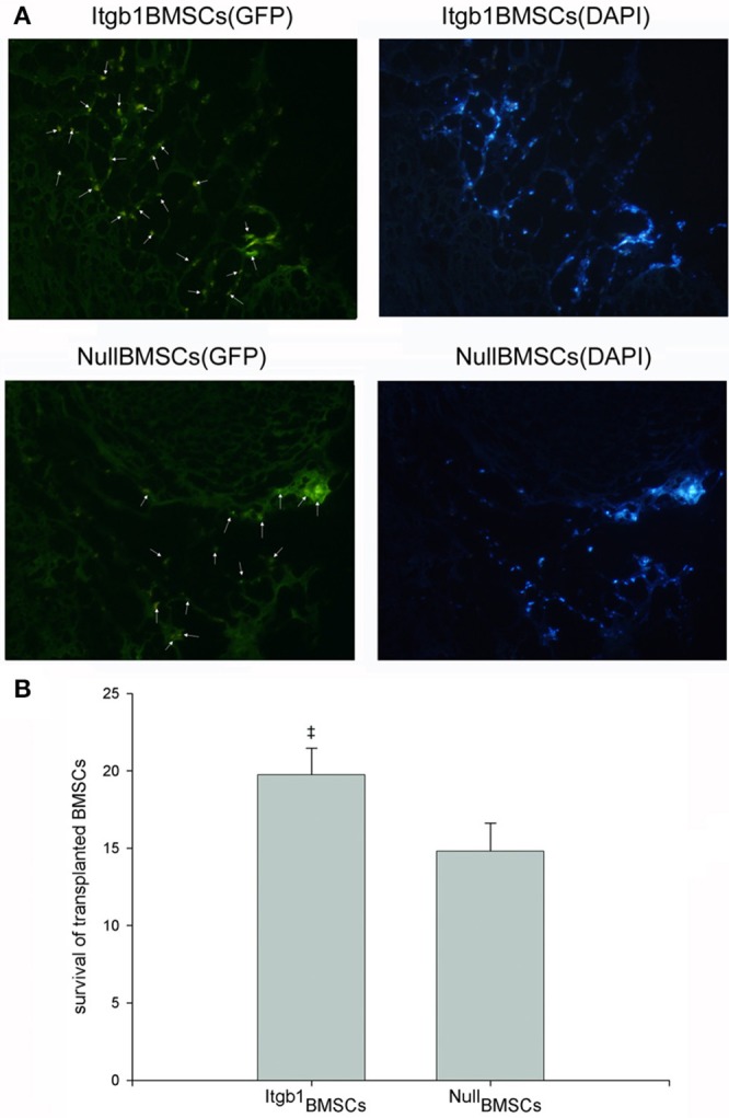
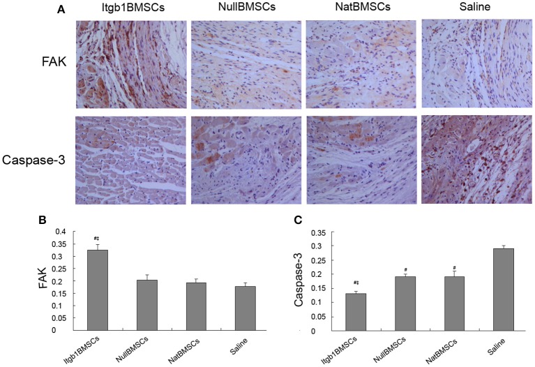
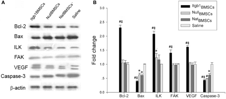
Bone mesenchymal stem cells (BMSCs) transplantation is a promising therapeutic approach for myocardial infarction (MI), but its application is limited by poor viability of BMSCs. In this study, we aimed to improve the survival of BMSCs by lentivirus vector mediated overexpression of integrin β1. In vitro study showed that integrin β1 overexpression could facilitate the proliferation of BMSCs under oxygen glucose deprivation condition and regulated the expression of Caspase-3, Bax, Bcl-2, FAK, and ILK in BMSCs. Next, MI was induced in rat model and Igtb1BMSCs, NullBMSCs, or NatBMSCs were transplanted by intramyocardial injection. One week later, the survival of BMSCs was higher in Itgb1 BMSCs group than in other groups. Four weeks after transplantation, heart function was significantly improved in Igtb1BMSCs group compared to other groups. The expression levels of Caspase-3 and Bax were decreased while the expression levels of Bcl-2, FAK, ILK, and VEGF were increased in the cardiomyocytes of Igtb1BMSCs group compared to other groups. In conclusion, integrin β1 overexpression could increase the survival of BMSCs and improve the efficacy of transplanted BMSCs for MI treatment. The beneficial effects may be mediated by inhibiting the apoptosis of both transplanted BMSCs and cardiomyocytes through adhesion-mediated cell survival signaling.
Keywords: adhesion; apoptosis; bone mesenchymal stem cell; integrin β1; myocardial infarction.
Figures
Similar articles
-
Catalpol Promotes the Survival and VEGF Secretion of Bone Marrow-Derived Stem Cells and Their Role in Myocardial Repair After Myocardial Infarction in Rats.Cardiovasc Toxicol. 2018 Oct;18(5):471-481. doi: 10.1007/s12012-018-9460-4. Cardiovasc Toxicol. 2018. PMID: 29752623
-
The harsh microenvironment in infarcted heart accelerates transplanted bone marrow mesenchymal stem cells injury: the role of injured cardiomyocytes-derived exosomes.Cell Death Dis. 2018 Mar 2;9(3):357. doi: 10.1038/s41419-018-0392-5. Cell Death Dis. 2018. PMID: 29500342 Free PMC article.
-
Edaravone promotes activation of resident cardiac stem cells by transplanted mesenchymal stem cells in a rat myocardial infarction model.J Thorac Cardiovasc Surg. 2016 Aug;152(2):570-82. doi: 10.1016/j.jtcvs.2016.02.071. Epub 2016 Mar 12. J Thorac Cardiovasc Surg. 2016. PMID: 27056755
-
CD51 distinguishes a subpopulation of bone marrow mesenchymal stem cells with distinct migratory potential: a novel cell-based strategy to treat acute myocardial infarction in mice.Stem Cell Res Ther. 2019 Nov 20;10(1):331. doi: 10.1186/s13287-019-1439-y. Stem Cell Res Ther. 2019. PMID: 31747966 Free PMC article.
-
A brief review: the therapeutic potential of bone marrow mesenchymal stem cells in myocardial infarction.Stem Cell Res Ther. 2017 Nov 2;8(1):242. doi: 10.1186/s13287-017-0697-9. Stem Cell Res Ther. 2017. PMID: 29096705 Free PMC article. Review.
Cited by
-
Global research trends and emerging opportunities for integrin adhesion complexes in cardiac repair: a scientometric analysis.Front Cardiovasc Med. 2024 Apr 18;11:1308763. doi: 10.3389/fcvm.2024.1308763. eCollection 2024. Front Cardiovasc Med. 2024. PMID: 38699584 Free PMC article.
-
Injectable Stem Cell-Based Therapies for Myocardial Regeneration: A Review of the Literature.J Funct Biomater. 2025 Apr 23;16(5):152. doi: 10.3390/jfb16050152. J Funct Biomater. 2025. PMID: 40422817 Free PMC article. Review.
-
Scalable Generation of Nanovesicles from Human-Induced Pluripotent Stem Cells for Cardiac Repair.Int J Mol Sci. 2022 Nov 18;23(22):14334. doi: 10.3390/ijms232214334. Int J Mol Sci. 2022. PMID: 36430812 Free PMC article.
-
Recent Progress in Stem Cell Modification for Cardiac Regeneration.Stem Cells Int. 2018 Jan 16;2018:1909346. doi: 10.1155/2018/1909346. eCollection 2018. Stem Cells Int. 2018. PMID: 29535769 Free PMC article. Review.
-
The role of integrin family in bone metabolism and tumor bone metastasis.Cell Death Discov. 2023 Apr 10;9(1):119. doi: 10.1038/s41420-023-01417-x. Cell Death Discov. 2023. PMID: 37037822 Free PMC article. Review.
References
-
- Bartunek J., Terzic A., Davison B. A., Filippatos G. S., Radovanovic S., Beleslin B., et al. . (2016). Cardiopoietic cell therapy for advanced ischemic heart failure: results at 39 weeks of the prospective, randomized, double blind, sham-controlled CHART-1 clinical trial. Eur. Heart J. 23:ehw543. 10.1093/eurheartj/ehw543 - DOI - PMC - PubMed
LinkOut - more resources
Full Text Sources
Other Literature Sources
Molecular Biology Databases
Research Materials
Miscellaneous