. 2017 Mar 17:8:398.
doi: 10.3389/fmicb.2017.00398. eCollection 2017.

Transcriptomics Indicates Active and Passive Metronidazole Resistance Mechanisms in Three Seminal Giardia Lines

Affiliations

Transcriptomics Indicates Active and Passive Metronidazole Resistance Mechanisms in Three Seminal Giardia Lines

Brendan R E Ansell et al. Front Microbiol. .

Abstract

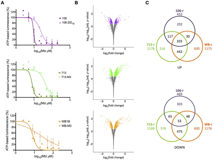
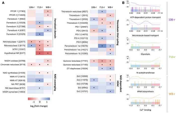
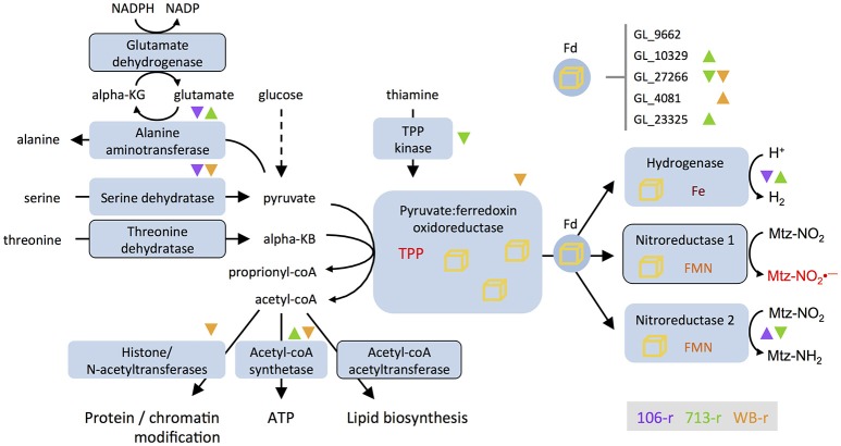
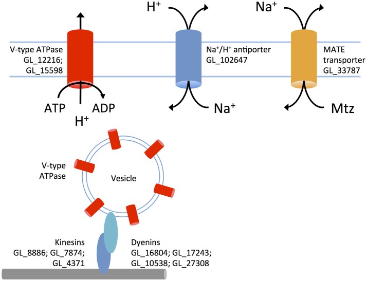
Giardia duodenalis is an intestinal parasite that causes 200-300 million episodes of diarrhoea annually. Metronidazole (Mtz) is a front-line anti-giardial, but treatment failure is common and clinical resistance has been demonstrated. Mtz is thought to be activated within the parasite by oxidoreductase enzymes, and to kill by causing oxidative damage. In G. duodenalis, Mtz resistance involves active and passive mechanisms. Relatively low activity of iron-sulfur binding proteins, namely pyruvate:ferredoxin oxidoreductase (PFOR), ferredoxins, and nitroreductase-1, enable resistant cells to passively avoid Mtz activation. Additionally, low expression of oxygen-detoxification enzymes can allow passive (non-enzymatic) Mtz detoxification via futile redox cycling. In contrast, active resistance mechanisms include complete enzymatic detoxification of the pro-drug by nitroreductase-2 and enhanced repair of oxidized biomolecules via thioredoxin-dependent antioxidant enzymes. Molecular resistance mechanisms may be largely founded on reversible transcriptional changes, as some resistant lines revert to drug sensitivity during drug-free culture in vitro, or passage through the life cycle. To comprehensively characterize these changes, we undertook strand-specific RNA sequencing of three laboratory-derived Mtz-resistant lines, 106-2ID10, 713-M3, and WB-M3, and compared transcription relative to their susceptible parents. Common up-regulated genes encoded variant-specific surface proteins (VSPs), a high cysteine membrane protein, calcium and zinc channels, a Mad-2 cell cycle regulator and a putative fatty acid α-oxidase. Down-regulated genes included nitroreductase-1, putative chromate and quinone reductases, and numerous genes that act proximal to PFOR. Transcriptional changes in 106-2ID10 diverged from those in 713-r and WB-r (r ≤ 0.2), which were more similar to each other (r = 0.47). In 106-2ID10, a nonsense mutation in nitroreductase-1 transcripts could enhance passive resistance whereas increased transcription of nitroreductase-2, and a MATE transmembrane pump system, suggest active drug detoxification and efflux, respectively. By contrast, transcriptional changes in 713-M3 and WB-M3 indicated a higher oxidative stress load, attributed to Mtz- and oxygen-derived radicals, respectively. Quantitative comparisons of orthologous gene transcription between Mtz-resistant G. duodenalis and Trichomonas vaginalis, a closely related parasite, revealed changes in transcripts encoding peroxidases, heat shock proteins, and FMN-binding oxidoreductases, as prominent correlates of resistance. This work provides deep insight into Mtz-resistant G. duodenalis, and illuminates resistance-associated features across parasitic species.

Keywords: Giardia; RNA sequencing (RNA-Seq); Trichomonas; messenger RNA; metronidazole resistance; oxidoreductases; single nucleotide polymorphism.


Figures

Figure 1
Figure 1
Metronidazole dose-response and differential transcription in resistant lines. (A) Dose-response curves for metronidazole-susceptible lines (dark colors) and resistant progeny (light colors). Experiments performed in biological duplicate. Error bars represent ± 1 standard deviation. (B) Fold-change in transcriptional abundance in resistant lines relative to susceptible parents. Significantly differentially transcribed genes (corrected p < 0.01) are displayed in color. Y-axes represent log-log-transformed corrected p values, where higher numbers indicate greater significance. (C) Numbers of significantly up-regulated (top panel) and down-regulated (bottom panel) genes shared between resistant lines. The total number of differentially transcribed genes is displayed beneath the line name.
Figure 2
Figure 2
Transcriptional changes in oxidoreductase-coding genes and related genes. (A) Color-scaled log2(fold change) values for resistant lines. Black dots indicate a statistically significant difference. Genes are grouped based on structure and/or function. Accession numbers (prefix: GL50803) appear in brackets. (B) Bar-code plots indicating the rank and log2(fold change) of genes (high: left, to low: right) comprising selected gene sets, below curves displaying sliding enrichment ratios. Blue and red shading indicate upper and lower quartiles, respectively.
Figure 3
Figure 3
Differential transcription of genes involved in ferredoxin-based electron transport and related processes. Nitroreductases are depicted as substrates of ferredoxin (Fd), as postulated by Ali and Nozaki (2007). The glutamate shunt consumes NADPH and cycles glutamate and α-ketoglutarate to drive the conversion of pyruvate to alanine. Enzymes encoded by genes that are down-regulated in all three resistant lines are outlined. Direction of differential transcription in different lines is indicated with color-coded up- or down-pointing triangles. Differential transcription for five ferredoxin-coding genes appears at top-right. Iron sulfur clusters are depicted as yellow cubes; Nitroreductase-1 activates metronidazole to toxic intermediates (Mtz-NO2, red). Nitroreductase-2 reduces Mtz to an inert amine (Mtz-NH2). KG, ketoglutarate; KB, ketobutyrate; coA, co-enzyme A; TPP, thiamine pyrophosphate; FMN, flavin mononucleotide; Fe, iron.
Figure 4
Figure 4
A putative metronidazole efflux system in 106-r. V-type ATPases are inserted into vesicular membranes and transported via microtubules and dynein/kinesin motors to the plasma membrane. These pumps use ATP to transport protons out of the cytosol against the diffusion gradient. An up-regulated sodium/proton antiporter exchanges in-flowing protons for sodium, increasing the extracellular sodium concentration. The MATE transporter is proposed to expel metronidazole as sodium flows back into the cell. Up-regulated genes (prefix GL50803 abbreviated to GL) are displayed next to proteins.
Figure 5
Figure 5
Putative structures for FMN-dependent quinone reductases and transcriptional regulation. (A) Transcripts encoding a quinone-reductase like enzyme (GL_17151) are down-regulated in all resistant lines. Predicted structures for this enzyme and two paralogs are shown at bottom. The three predicted protein stuctures are similar to the crystal structure of KefF (PDB code 3EYW), displayed at centre. Monomers within dimers forming the Kef complex are coloured dark blue (KefF), and light blue (KefC). Two proteins with a KefC-like fold were identified (at top), of which GL_8468 shows a transcriptional profile similar to GL_17150. Differential transcription of genes encoding these proteins is indicated with color-coded up- or down-pointing triangles (purple, 106-r; green, 713-r; orange, WB-r). (B) Sense (top panel) and antisense (bottom panel) transcript abundance over a region in chromosome 5 encoding the paralogous putative quinone reductases, GL_17151 and GL_17150. Normalized read depth for three replicates of 713-s (gray lines) and 713-r (green lines) is displayed, and is representative of other lines. Gene models are displayed in black (forward strand) and gray (reverse strand). (C) Pearson correlations of log2(fold change) values for sense (x-axis) and antisense (y-axis) transcripts in metronidazole-resistant lines, relative to susceptible parent lines. Positive correlations for 106-r and 713-r are displayed in red.

Similar articles

Cited by

References

    1. Adagu I. S., Nolder D., Warhurst D. C., Rossignol J.-F. (2001). In vitro activity of nitazoxanide and related compounds against isolates of Giardia intestinalis, Entamoeba histolytica and Trichomonas vaginalis. J. Antimicrob. Chemother. 49, 103–111. 10.1093/jac/49.1.103 - DOI - PubMed
    1. Adam R. D., Nigam A., Seshadri V., Martens C. A., Farneth G. A., Morrison H. G., et al. . (2010). The Giardia lamblia vsp gene repertoire: characteristics, genomic organization, and evolution. BMC Genomics 11:424. 10.1186/1471-2164-11-424 - DOI - PMC - PubMed
    1. Ali V., Nozaki T. (2007). Current therapeutics, their problems, and sulfur-containing-amino-acid metabolism as a novel target against infections by “amitochondriate” protozoan parasites. Clin. Microbiol. Rev. 20, 164–187. 10.1128/CMR.00019-06 - DOI - PMC - PubMed
    1. Ansell B. R. E., McConville M. J., Baker L., Korhonen P. K., Emery S. J., Svärd S. G., et al. . (2016). Divergent transcriptional responses to physiological and xenobiotic stress in Giardia duodenalis. Antimicrob. Agents Chemother. 60, AAC.00977–00916. 10.1128/AAC.00977-16 - DOI - PMC - PubMed
    1. Ansell B. R. E., McConville M. J., Baker L., Korhonen P. K., Young N. D., Hall R. S., et al. . (2015a). Time-dependent transcriptional changes in axenic Giardia duodenalis trophozoites. PLoS Negl. Trop. Dis. 9:e0004261. 10.1371/journal.pntd.0004261.s013 - DOI - PMC - PubMed

LinkOut - more resources