. 2016 Aug;11(4):261-284.
doi: 10.1007/s11515-016-1407-1. Epub 2016 Jul 8.

Neural stem cell heterogeneity through time and space in the ventricular-subventricular zone

Affiliations

Neural stem cell heterogeneity through time and space in the ventricular-subventricular zone

Gabrielle Rushing et al. Front Biol (Beijing). 2016 Aug.

Abstract

Background: The origin and classification of neural stem cells (NSCs) has been a subject of intense investigation for the past two decades. Efforts to categorize NSCs based on their location, function and expression have established that these cells are a heterogeneous pool in both the embryonic and adult brain. The discovery and additional characterization of adult NSCs has introduced the possibility of using these cells as a source for neuronal and glial replacement following injury or disease. To understand how one could manipulate NSC developmental programs for therapeutic use, additional work is needed to elucidate how NSCs are programmed and how signals during development are interpreted to determine cell fate.

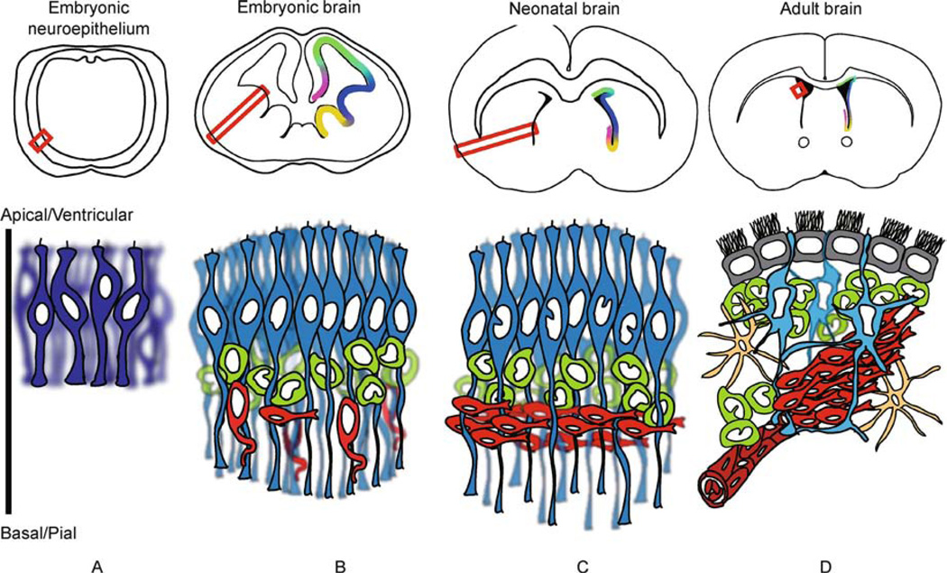
Objective: This review describes the identification, classification and characterization of NSCs within the large neurogenic niche of the ventricular-subventricular zone (V-SVZ).

Methods: A literature search was conducted using Pubmed including the keywords "ventricular-subventricular zone," "neural stem cell," "heterogeneity," "identity" and/or "single cell" to find relevant manuscripts to include within the review. A special focus was placed on more recent findings using single-cell level analyses on neural stem cells within their niche(s).

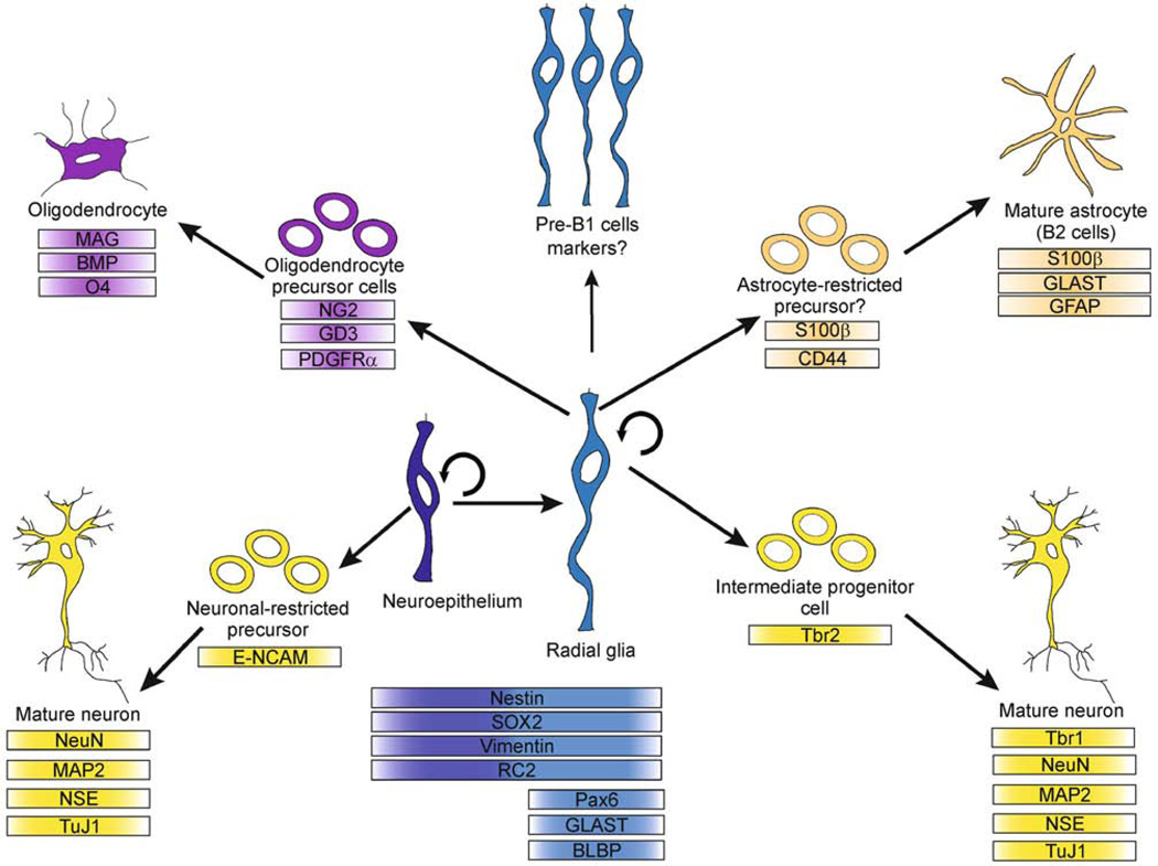
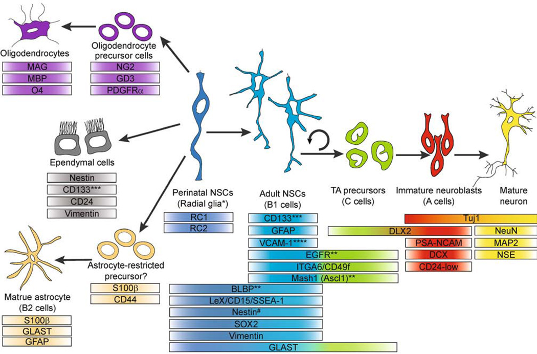
Results: This review discusses over 20 research articles detailing findings on V-SVZ NSC heterogeneity, over 25 articles describing fate determinants of NSCs, and focuses on 8 recent publications using distinct single-cell analyses of neural stem cells including flow cytometry and RNA-seq. Additionally, over 60 manuscripts highlighting the markers expressed on cells within the NSC lineage are included in a chart divided by cell type.

Conclusions: Investigation of NSC heterogeneity and fate decisions is ongoing. Thus far, much research has been conducted in mice however, findings in human and other mammalian species are also discussed here. Implications of NSC heterogeneity established in the embryo for the properties of NSCs in the adult brain are explored, including how these cells may be redirected after injury or genetic manipulation.

Keywords: heterogeneity; neural stem cells; positional identity; single-cell; ventricular-subventricular zone.


Conflict of interest statement

The authors declare that they have no conflicts of interest.

Figures

Figure 1
Figure 1
Development of the mouse ventricular-subventricular zone. Representative coronal sections of mouse brain at indicated developmental times are shown at top. Colors in the coronal sections represent domains of transcription factor expression within the developing V-SVZ. At bottom, representative schematics of developing V-SVZ corresponding to red box within the coronal section above. Note that the size of coronal sections and corresponding representative images of cell types are not to scale. (A) Neuroepithelial cells (NECs; dark blue) fold in to form the neural tube. These cells contact both the pial and ventricular surfaces of the developing brain (below) and divide to form a densely packed VZ. (B) In developing telencephalon, NECs give rise to radial glia (RG; light blue), which retain properties of NECs (see text), including contact with the ventricular and pial surfaces. At this stage, the RG divide asymmetrically, producing a daughter RG and a daughter intermediate progenitor cell (IPC; green) located away from the ventricular surface in a subventricular zone (SVZ). Newborn neurons (red) use the RG processes as a scaffold for migration to their final destinations. (C) In the neonatal brain, the RG are retained until approximately postnatal day 7. After postnatal day 2, they begin to retract their basal (pial) processes and will give rise to ependymal (E) cells (grey; shown in (D)), B1 cells (teal; shown in (D)) and B2 cells (yellow; shown in (D)) in the mature brain. (D) In the adult brain, B1 cells are the NSCs. They have basal processes that wrap around blood vessels (dark red) and a single primary cilium that extends between the tightly connected E cells. The multiple motile cilia of E cells push CSF through the ventricles. Note the presence of transit-amplifying C cells (green), migrating neuroblasts (A cells, red) and parenchymal astrocytes (B2 cells, orange). This structure in the adult is termed the ventricular-subventricular zone (V-SVZ). Note that other cell types exist in the region that are not discussed within this review including microglia and local and distant innervating neurons.
Figure 2
Figure 2
Embryonic neural cell lineage and marker expression profiles. Neuroepithelial cells (NECs) are the earliest neural progenitors discussed here. These cells produce neurons but also give rise to radial glia cells (RG), which in turn act as the primary progenitors during cortical development. These cells can produce oligodendrocytes, astrocytes and neurons. While precursors for oligodendrocytes and neurons have been characterized, it is still debated whether an astrocyte-restricted precursor cell exists. RG cells also produce pre-B1 cells between E13.5–15.5 that remain relatively quiescent until postnatal reactivation (see text).
Figure 3
Figure 3
Postnatal neural cell lineage and marker expression profiles. *Radial glia persist only during the first postnatal week and are non-self-renewing during this time. They retract their processes after postnatal day 2 in the mouse and give rise to parenchymal astrocytes (orange), ependymal cells (grey), oligodendrocytes (purple) and astrocyte-like adult neural stem cells (B1 cells, teal). B1 cells are self-renewing and also give rise to transit amplifying progenitors (green), which in turn produce neuroblasts (red) that will mature into neurons (yellow). **These markers are primarily expressed by activated B1 cells. ***CD133 is present on the primary cilia of B1 cells, as well as ependymal cells. ****VCAM-1 is expressed on quiescent B1 cells. # Note that Nestin is not expressed on all RG and B1 cells but rather, is dynamically regulated (see text).

Similar articles

Cited by

References

    1. Aguirre A, Rubio ME, Gallo V. Notch and EGFR pathway interaction regulates neural stem cell number and self-renewal. Nature. 2010;467(7313):323–327. - PMC - PubMed
    1. Ahn S, Joyner AL. In vivo analysis of quiescent adult neural stem cells responding to Sonic hedgehog. Nature. 2005;437(7060):894–897. - PubMed
    1. Altman J. Autoradiographic study of degenerative and regenerative proliferation of neuroglia cells with tritiated thymidine. Exp Neurol. 1962;5(4):302–318. - PubMed
    1. Altman J, Das GD. Autoradiographic and histological evidence of postnatal hippocampal neurogenesis in rats. J Comp Neurol. 1965;124(3):319–335. - PubMed
    1. Alvarez-Buylla A, García-Verdugo JM, Tramontin AD. A unified hypothesis on the lineage of neural stem cells. Nat Rev Neurosci. 2001;2(4):287–293. - PubMed

LinkOut - more resources