. 2017:2017:4128061.
doi: 10.1155/2017/4128061. Epub 2017 Mar 7.

Spermidine Oxidation-Mediated Degeneration of Retinal Pigment Epithelium in Rats

Affiliations

Spermidine Oxidation-Mediated Degeneration of Retinal Pigment Epithelium in Rats

Koji Ohashi et al. Oxid Med Cell Longev. 2017.

Abstract

Retinal pigment epithelium (RPE) degeneration is a crucial event in dry age-related macular degeneration and gyrate atrophy. The polyamine spermidine has been shown to induce RPE cell death in vitro. The present study aimed to establish a novel in vivo model of spermidine-induced RPE degeneration and to determine whether spermidine-induced RPE cell death involves oxidative mechanisms. In this study, spermidine caused ARPE-19 cell death in a concentration-dependent manner. This effect was prevented by removal of serum from the culture medium or treatment with amine oxidase inhibitors, N-acetylcysteine (NAC), or aldehyde dehydrogenase (ALDH). Intravitreal injection of spermidine into rats significantly increased the permeability of the blood-retinal barrier and decreased the amplitudes of scotopic electroretinogram a- and b-waves. Histological analysis revealed that spermidine induced vacuolation, atrophy, and dropout of RPE cells, leading to the disruption of photoreceptor outer segments. Simultaneous intravitreal administration of NAC and ALDH with spermidine prominently inhibited the functional and morphological changes induced by spermidine. In conclusion, this study demonstrated that the intravitreal administration of spermidine induced RPE cell dysfunction and death followed by photoreceptor degeneration in rats. These effects of spermidine are thought to be mediated by oxidative stress and a toxic aldehyde generated during spermidine oxidation.


Conflict of interest statement

The authors declare that there is no conflict of interests regarding the publication of this paper.

Figures

Figure 1
Figure 1
Effects of serum removal and treatment with amine oxidase inhibitors, an antioxidant compound, and aldehyde dehydrogenase (ALDH) on spermidine-induced ARPE-19 cell death. Cell viability was assessed with a MTS assay 24 h after the addition of spermidine. The effects of (a) serum removal and treatment with (b) amine oxidase inhibitors, (c) an antioxidant, and (d) ALDH when applied simultaneously with spermidine. Each column represents a mean ± standard error of the mean of four wells. ##P < 0.01 versus control. ∗∗P < 0.01 versus spermidine. AG: aminoguanidine. PEN: pentamidine.
Figure 2
Figure 2
Spermidine-induced hyperpermeability of the blood-retinal barrier (BRB) in rats. BRB permeability was assessed by vitreous fluorophotometry on days 1, 3, 7, 14, and 28 after the intravitreal administration of spermidine. Each value represents the mean ± standard error of the mean of six to eight eyes. P < 0.05, ∗∗P < 0.01 versus control.
Figure 3
Figure 3
Spermidine-induced impairment of electroretinogram (ERG) a- and b-wave amplitudes in rats. Scotopic ERG a- and b-waves were measured on days 2, 6, 13, and 27 days after the intravitreal administration of spermidine. Traces typical of (a) control, (b) spermidine (10 nmol/eye), (c) spermidine (20 nmol/eye), and (d) spermidine (30 nmol/eye) treatments, 13 days after the injection are shown. Time course data of (e) ERG a-waves and (f) ERG b-waves are shown. Each value represents the mean ± standard error of the mean of seven to eight eyes. P < 0.05 and ∗∗P < 0.01 versus control.
Figure 4
Figure 4
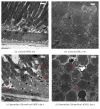
Spermidine-induced degeneration of retinal pigment epithelium (RPE) and photoreceptor cells in rats. Cross-sections of rat eyes were prepared (a, b) 1 day after the administration of DPBS, (c–l) 1, 3, 7, 14, and 28 days after the intravitreal administration of spermidine (20 nmol/eye), and (m–o) 7 days after the injection of spermidine (10 or 30 nmol/eye). Black and white scale bars indicate 50 and 20 μm, respectively. Red arrows in (d) indicate RPE cell vacuolization. Red arrows in (f), (h), and (p) indicate RPE cell degeneration. Red arrows in (j) and (l) indicate RPE cell regeneration. Yellow arrows in (h), (j), (l), and (p) indicate photoreceptor cell degeneration. NFL-GCL: nerve fiber layer and ganglion cell layer. IPL: inner plexiform layer. INL: inner nuclear layer. OPL: outer plexiform layer. ONL: outer nuclear layer. IS: inner segment. OS: outer segment. RPE: retinal pigment epithelium. C: choroid.
Figure 5
Figure 5
Spermidine-induced ultrastructural changes in the retinal pigment epithelium (RPE) and outer nuclear layer (ONL) of the rat retina. Ultrathin 80–nm sections were prepared from samples fixed 6 hours after the injection of (a, b) DPBS or 4 days after the intravitreal administration of (c, d) spermidine (20 nmol/eye). Scale bars indicate 2 μm. Red arrows in (c) indicate plasma membrane disruption in RPE cells. Red arrows in (d) indicate chromatin condensation in photoreceptor cells.
Figure 6
Figure 6
Protective effects of N-acetylcysteine (NAC) and aldehyde dehydrogenase (ALDH) on spermidine-induced hyperpermeability of the blood-retinal barrier (BRB). (a) BRB permeability was assessed by vitreous fluorophotometry 7 days after the intravitreal administration of (a) spermidine (20 nmol/eye) alone, (b) spermidine (20 nmol/eye) plus NAC (500 nmol/eye), and (c) spermidine (20 nmol/eye) plus ALDH (1.5 U/eye). Each column represents a mean ± standard error of the mean of seven to eight eyes. ##P < 0.01 versus control and P < 0.05 and ∗∗P < 0.01 versus spermidine (20 nmol/eye).
Figure 7
Figure 7
Protective effects of N-acetylcysteine (NAC) and aldehyde dehydrogenase (ALDH) on spermidine-induced impairment of electroretinogram (ERG) a- and b-wave amplitudes. Representative traces are shown of scotopic ERG a- and b-waves measured 13 days after the administration of (a) control, (b) spermidine (20 nmol/eye) alone, (c) spermidine (20 nmol/eye) plus NAC (500 nmol/eye), and (d) spermidine (20 nmol/eye) plus ALDH (1.5 U/eye). The amplitude data for ERG a- and b-waves, from eyes treated as above are depicted in (e) and (f), respectively. Each column represents the mean ± standard error of the mean of seven to eight eyes. ##P < 0.01 versus control and ∗∗P < 0.01 versus spermidine (20 nmol/eye).
Figure 8
Figure 8
Protective effects of N-acetylcysteine (NAC) and aldehyde dehydrogenase (ALDH) on spermidine-induced degeneration of the retinal pigment epithelium (RPE) and photoreceptors in rats. Cross-sections of rat eyes were prepared 13 days after the intravitreal injection of (a, b) spermidine (20 nmol/eye) alone, (c, d) spermidine (20 nmol/eye) plus NAC (500 nmol/eye), and (e, f) spermidine (20 nmol/eye) plus ALDH (1.5 U/eye). Black and white scale bars indicate 50 and 20 μm, respectively. NFL-GCL: nerve fiber layer and ganglion cell layer. IPL: inner plexiform layer. INL: inner nuclear layer. OPL: outer plexiform layer. ONL: outer nuclear layer. IS-OS-RPE: inner segment, outer segment, and retinal pigment epithelium. C: choroid.
Figure 9
Figure 9
Immunohistochemistry of acrolein in spermidine-treated rat retina. Immunohistochemical localization of acrolein was performed using cross-sections of rat eyes which were prepared (a) 1 day and (b) 3 days after the intravitreal injection of DPBS or spermidine (20 nmol/eye). Scale bars indicate 20 μm. Inserted photograph in (b) is an enlarged image of the same slide.

References

    1. Strauss O. The retinal pigment epithelium in visual function. Physiological Reviews. 2005;85(3):845–881. doi: 10.1152/physrev.00021.2004. - DOI - PubMed
    1. Ambati J., Fowler B. J. Mechanisms of age-related macular degeneration. Neuron. 2012;75(1):26–39. doi: 10.1016/j.neuron.2012.06.018. - DOI - PMC - PubMed
    1. Green W. R., Enger C. Age-related macular degeneration histopathologic studies: the 1992 Lorenz E. Zimmerman lecture. Ophthalmology. 1993;100(10):1519–1535. doi: 10.1016/s0161-6420(93)31466-1. - DOI - PubMed
    1. Wilson D. J., Weleber R. G., Green W. R. Ocular clinicopathologic study of gyrate atrophy. American Journal of Ophthalmology. 1991;111(1):24–33. doi: 10.1016/S0002-9394(14)76892-8. - DOI - PubMed
    1. Bowes Rickman C., Farsiu S., Toth C. A., Klingeborn M. Dry age-related macular degeneration: mechanisms, therapeutic targets, and imaging. Investigative Ophthalmology and Visual Science. 2013;54(14):ORSF68–ORSF80. doi: 10.1167/iovs.13-12757. - DOI - PMC - PubMed

MeSH terms

LinkOut - more resources