. 2017 Mar 29:8:17.
doi: 10.1186/s13229-017-0134-z. eCollection 2017.

Transcriptome analysis of microglia in a mouse model of Rett syndrome: differential expression of genes associated with microglia/macrophage activation and cellular stress

Affiliations

Transcriptome analysis of microglia in a mouse model of Rett syndrome: differential expression of genes associated with microglia/macrophage activation and cellular stress

Dejian Zhao et al. Mol Autism. .

Abstract

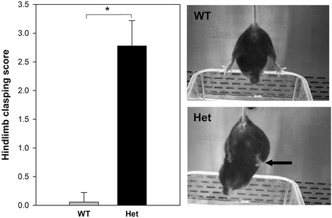
Background: Rett syndrome (RTT) is a severe, neurodevelopmental disorder primarily affecting girls, characterized by progressive loss of cognitive, social, and motor skills after a relatively brief period of typical development. It is usually due to de novo loss of function mutations in the X-linked gene, MeCP2, which codes for the gene expression and chromatin regulator, methyl-CpG binding protein 2. Although the behavioral phenotype appears to be primarily due to neuronal Mecp2 deficiency in mice, other cell types, including astrocytes and oligodendrocytes, also appear to contribute to some aspects of the RTT phenotype. In addition, microglia may also play a role. However, the effect of Mecp2 deficiency in microglia on RTT pathogenesis is controversial.

Methods: In the current study, we applied whole transcriptome analysis using RNA-seq to gain insight into molecular pathways in microglia that might be dysregulated during the transition, in female mice heterozygous for an Mecp2-null allele (Mecp2+/-; Het), from the pre-phenotypic (5 weeks) to the phenotypic phases (24 weeks).

Results: We found a significant overlap in differentially expressed genes (DEGs) with genes involved in regulating the extracellular matrix, and those that are activated or inhibited when macrophages and microglia are stimulated towards the M1 and M2 activation states. However, the M1- and M2-associated genes were different in the 5- and 24-week samples. In addition, a substantial decrease in the expression of nine members of the heat shock protein (HSP) family was found in the 5-week samples, but not at 24 weeks.

Conclusions: These findings suggest that microglia from pre-phenotypic and phenotypic female mice are activated in a manner different from controls and that pre-phenotypic female mice may have alterations in their capacity to response to heat stress and other stressors that function through the HSP pathway.

Keywords: Autism; Heat shock; Innate immune system; M1 activation; M2 activation; Microglia; Rett syndrome; Schizophrenia.


Figures

Fig. 1
Fig. 1
Left panel. Hindlimb clasping score. Het female mice were tested biweekly for the hindlimb clasping reflex and scored on a scale of 0–3 as described in the Methods section. All mice began to show the reflex after week 23. They were then scored over 3 consecutive days, and the results for each mouse were pooled. There was a significant increase in the score in the Het mice (denoted by asterisk, 7.4E−08, Student’s t test). Right upper panel WT mice consistently showed a lack of hindlimb clasping (outward hindlimbs) while the Het mice showed an abnormal response; clasping towards the abdomen (arrow  Right lower panel)
Fig. 2
Fig. 2
Heat map and summary of GO terms and pathways. a, c Heat maps showing differentially expressed genes in microglia between Het and WT at 5 and 24 weeks. b, d Enriched GO terms and pathways determined by the software ToppGene. The terms shown are the top enrichment terms for various categories in the DEG lists. These included gene ontology (GO) (molecular function [MF] and biological process [BP]), mouse phenotype, protein domain, co-expression, and PubMed references. All enriched terms are included in Additional file 6: Table S3
Fig. 3
Fig. 3
Validation of selected DEGs by qPCR. Samples were analyzed in duplicate using the 2−∆∆Ct method as described in the Methods section. Significant differences between WT and Het are denoted by an asterisk (*). p values were derived by Student’s t test. The values are as follows: 5-week CD11b-positive fraction (microglia); Hspa8, 0.01; Hspa1a, 0.02; Hspa1b, 0.005; Hsph1, 0.08; Dnaja1, 0.2; Mecp2, 0.03, CD180, 0.03: 5-week CD11b-negative fraction; Hspa8, 0.03; Hspa1a, 0.2; Hspa1b, 0.01; Hsph1, 0.9; Dnaja1, 0.9; Mecp2, 0.01, CD180, 0.006: 24-week CD11b-positive fraction: Hspa8, 0.09; Hspa1a, 0.5; Hspa1b, 0.1; Hsph1, 0.4; Dnaja1, 0.09; Mecp2, 0.0001, CD180, 0.2: 24-week CD11b negative fraction; Hspa8, 0.1; Hspa1a, 0.9; Hspa1b, not done; Hsph1, 0.1; Dnaja1, 0.7; Mecp2, 0.1; CD180, 0.3

Similar articles

Cited by

References

    1. Amir RE, Van den Veyver IB, Wan M, Tran CQ, Francke U, Zoghbi HY. Rett syndrome is caused by mutations in X-linked MECP2, encoding methyl-CpG-binding protein 2. Nat Genet. 1999;23(2):185–8. doi: 10.1038/13810. - DOI - PubMed
    1. Pohodich AE, Zoghbi HY. Rett syndrome: disruption of epigenetic control of postnatal neurological functions. Hum Mol Genet. 2015;24(R1):R10–6. doi: 10.1093/hmg/ddv217. - DOI - PMC - PubMed
    1. Mellen M, Ayata P, Dewell S, Kriaucionis S, Heintz N. MeCP2 binds to 5hmC enriched within active genes and accessible chromatin in the nervous system. Cell. 2012;151(7):1417–30. doi: 10.1016/j.cell.2012.11.022. - DOI - PMC - PubMed
    1. Guo JU, Su Y, Shin JH, Shin J, Li H, Xie B, Zhong C, Hu S, Le T, Fan G, Zhu H, Chang Q, Gao Y, Ming GL, Song H. Distribution, recognition and regulation of non-CpG methylation in the adult mammalian brain. Nat Neurosci. 2014;17(2):215–22. doi: 10.1038/nn.3607. - DOI - PMC - PubMed
    1. Cheng TL, Wang Z, Liao Q, Zhu Y, Zhou WH, Xu W, Qiu Z. MeCP2 suppresses nuclear microRNA processing and dendritic growth by regulating the DGCR8/Drosha complex. Dev Cell. 2014;28(5):547–60. doi: 10.1016/j.devcel.2014.01.032. - DOI - PubMed

Publication types