. 2017:2017:6468356.
doi: 10.1155/2017/6468356. Epub 2017 Mar 7.

Evidence of Presynaptic Localization and Function of the c-Jun N-Terminal Kinase

Affiliations

Evidence of Presynaptic Localization and Function of the c-Jun N-Terminal Kinase

Silvia Biggi et al. Neural Plast. 2017.

Abstract

The c-Jun N-terminal kinase (JNK) is part of a stress signalling pathway strongly activated by NMDA-stimulation and involved in synaptic plasticity. Many studies have been focused on the post-synaptic mechanism of JNK action, and less is known about JNK presynaptic localization and its physiological role at this site. Here we examined whether JNK is present at the presynaptic site and its activity after presynaptic NMDA receptors stimulation. By using N-SIM Structured Super Resolution Microscopy as well as biochemical approaches, we demonstrated that presynaptic fractions contained significant amount of JNK protein and its activated form. By means of modelling design, we found that JNK, via the JBD domain, acts as a physiological effector on T-SNARE proteins; then using biochemical approaches we demonstrated the interaction between Syntaxin-1-JNK, Syntaxin-2-JNK, and Snap25-JNK. In addition, taking advance of the specific JNK inhibitor peptide, D-JNKI1, we defined JNK action on the SNARE complex formation. Finally, electrophysiological recordings confirmed the role of JNK in the presynaptic modulation of vesicle release. These data suggest that JNK-dependent phosphorylation of T-SNARE proteins may have an important functional role in synaptic plasticity.


Conflict of interest statement

There is no actual or potential conflict of interests.

Figures

Figure 1
Figure 1
JNK is localized in the presynaptic compartment. (a) Immunofluorescence on mouse cortical purified synaptosomes stained for p-JNK (green) and for Syntaxin-2 (red) colocalization is visualized as yellow signal in Merge image, with Pearson's coefficient 0.54 ± 0.08. Images acquired with super resolution microscopy; scale bar 1 μm. (b) Western blotting analysis of JNK and p-JNK active form presence in 20 μg of total cortical lysates, postsynaptic density region (represented by PSD95 postsynaptic specific protein on TIF fraction) and presynaptic compartment (represented by Synaptophysin presynaptic specific protein on purified synaptosomes). JNK is localized both in postsynaptic and in presynaptic region and its phosphorylation state is comparable. Actin was used as loading control.
Figure 2
Figure 2
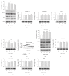
JNK is activated after pre-NMDARs stimulation in young mice and adult. (a–e) Western blotting and relative quantification of JNK activation (a), measured as p-JNK/JNK ratio, on p14 mice crude synaptosomes stimulated with NMDA 100 μM and glycine 1 μM for 2, 5, and 10 min. JNK activation significantly increases 2 min after treatment (p < 0.05) and persists at 5 min (p < 0.05). Western blotting and relative quantification of presynaptic proteins Syntaxin-1 (b), Syntaxin-2 (c), Snap25 (d), and Vamp (e) extracted from p14 mice crude synaptosomes stimulated with NMDA 100 μM and glycine 1 μM for 2, 5, and 10 min. Protein levels remain unchanged after treatment at the considered time-points. Actin was used as loading control (20 μg proteins loaded; N = 5). One-way ANOVA, Dunnet's post-hoc test. Data are showed as mean ± S.E.M. (f) Calcium currents, measured with calcium sensible fluorophore Fluo4, on crude synaptosomes treated with NMDA 100 μM and glycine 1 μM or with KCl 50 mM. Fluorescence was recorded with a spectrophotometric approach for 7 min, with NMDA + Gly or KCl injection at 45 sec. After both injections there was a significant increase in calcium levels starting from 45 sec after stimulation (each experimental group N = 5). One-way ANOVA, Dunnet's post hoc test. Data are shown as mean ± S.E.M. (g–k) Western blotting and relative quantification of JNK activation (g), measured as p-JNK/JNK ratio on adult mice crude synaptosomes stimulated with NMDA 100 μM and glycine 1 μM for 2, 5, and 10 min. JNK activation increases 2 min after treatment (∗∗∗p < 0.001) and persists at 5 min (∗∗∗p < 0.01) and at 10 min (∗∗p < 0.01). Western blotting and relative quantification of presynaptic proteins Syntaxin-1 (h), Syntaxin-2 (i), Snap25 (j), and Vamp (k) extracted from adult mice crude synaptosomes stimulated with NMDA 100 μM and glycine 1 μM for 2, 5, and 10 min. Protein levels remained unchanged after treatment for all three time-points. Actin was used as loading control (20 μg proteins loaded; N = 4). One-way ANOVA, Dunnet's post hoc test. Data are shown as mean ± S.E.M.
Figure 3
Figure 3
JBD is crucial for SNARE interaction with JNK. (a) Sequence alignment between Syntaxin-1 and Syntaxin-2. The JBDs are underlined in red. (b) Sequence of Snap25 with the JBD underlined in red. On top of panels (a) and (b) the canonical JBD pattern is also reported. (c) Best JNK-Syntaxin complex resulting from the docking predictions. JNK is shown in light blue and Syntaxin in gold. The lateral chains of the residues belonging to the JBD are shown in red. (d) Best complex of the JNK-Snap25 docking results. Also in this case JNK is depicted in light blue while Snap25 is shown in light brown. The lateral chains of the residues of the JBD in Snap25 are reported in red.
Figure 4
Figure 4
JNK preferentially interacts with Syntaxin-1, Syntaxin-2, and Snap25. (a–c) Western blotting after immunoprecipitation of Syntaxin-1 (a), Syntaxin-2 (b), and Snap25 (c) on purified synaptosomes lysates extracted from adult mice cortex. JNK is detectable in Syntaxin-1 and Syntaxin-2 precipitate, while immunoreactive signal is weak in Snap25 precipitate. (d) Both Syntaxin-1 and Syntaxin-2, as well as Snap25, were detectable in JNK precipitate. 500 μg of total lysates has been subjected to immunoprecipitation, while 20 μg was loaded as control. N = 4.
Figure 5
Figure 5
Both JNK2 and JNK3 interact with Syntaxin-2 and Snap25. Western blotting after immunoprecipitation of JNK2 (left) and JNK3 (right) on purified synaptosomes lysates extracted from adult mice cortex. Syntaxin-2 and Snap25 were detectable both in JNK2 and in JNK3 precipitate. 500 μg of total lysates has been subjected to immunoprecipitation, while 20 μg was loaded as control (each experimental group N = 3).
Figure 6
Figure 6
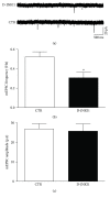
D-JNKI1 treatment reduced the frequency of mEPSCs in mice cortical slices. (a) Representative traces of mEPSCs recorded from cortical slices under control condition or after incubation with D-JNKI1n for 1 hr. (b-c) Bar graph (mean ± SEM) showing the mEPSC frequency (b) and amplitude (c) obtained in the first 5 min of recordings from control (N = 8) and D-JNKI1-treated (N = 8) cortical neurons. CTR versus D-JNKI1 treatment: p < 0.05 Student's t-test, n =8. Data are shown as mean +/− SEM.
Figure 7
Figure 7
JNK inhibition reduces SNARE complex formation. (a) Western blotting and relative quantification of SNARE complex levels (anti-Syntaxin-1 immunoreactive bands from approximately 75 to 100 KDa) in 20 μg of crude synaptosomes stimulated with NMDA 100 μm and glycine 1 μm for 7 min in presence or absence of D-JNKI1 (2 μm), added to synaptosomes 25 min before stimulation. After NMDA/Gly exposure there was a significant increase in SNARE complexes formation (p < 0.05), which is totally prevented by D-JNKI1 preadministration (#p < 0.05). SNARE complexes are normalized on actin, N = 6. Data are shown as mean ± SEM, Two-way ANOVA, Tukey's post hoc test. (b–d) Western blotting and relative quantification of Syntaxin-1, Syntaxin-2, and Snap25 in 20 μg of crude synaptosomes stimulated with NMDA 100 μm and glycine 1 μm for 7 min in presence or absence of D-JNKI1 (2 μm), added to synaptosomes 25 min before stimulation. There was no significant variation of protein levels after NMDA/Gly exposure as well as after D-JNKI1 administration alone or in combination with NMDA/Gly. Proteins are normalized on actin, N = 6. Two-way ANOVA, Tukey's post hoc test. Data are shown as mean ± SEM.

Similar articles

Cited by

References

    1. Davis R. J. Signal transduction by the JNK group of MAP kinases. Cell. 2000;103(2):239–252. doi: 10.1016/S0092-8674(00)00116-1. - DOI - PubMed
    1. Chang L., Karin M. Mammalian MAP kinase signalling cascades. Nature. 2001;410(6824):37–40. doi: 10.1038/35065000. - DOI - PubMed
    1. Angel P., Karin M. The role of Jun, Fos and the AP-1 complex in cell-proliferation and transformation. BBA—Reviews on Cancer. 1991;1072(2-3):129–157. doi: 10.1016/0304-419x(91)90011-9. - DOI - PubMed
    1. Kawauchi T., Chihama K., Nabeshima Y.-I., Hoshino M. The in vivo roles of STEF/Tiam1, Rac1 and JNK in cortical neuronal migration. EMBO Journal. 2003;22(16):4190–4201. doi: 10.1093/emboj/cdg413. - DOI - PMC - PubMed
    1. Bogoyevitch M. A., Kobe B. Uses for JNK: the many and varied substrates of the c-Jun N-terminal kinases. Microbiology and Molecular Biology Reviews. 2006;70(4):1061–1095. doi: 10.1128/mmbr.00025-06. - DOI - PMC - PubMed

Publication types

MeSH terms

Substances