. 2017:2017:5871043.
doi: 10.1155/2017/5871043. Epub 2017 Mar 7.

Synthetic Peptides as Potential Antigens for Cutaneous Leishmaniosis Diagnosis

Affiliations

Synthetic Peptides as Potential Antigens for Cutaneous Leishmaniosis Diagnosis

Juliana Seger Link et al. J Immunol Res. 2017.

Abstract

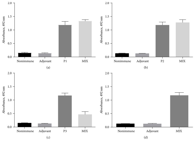
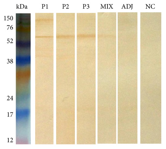
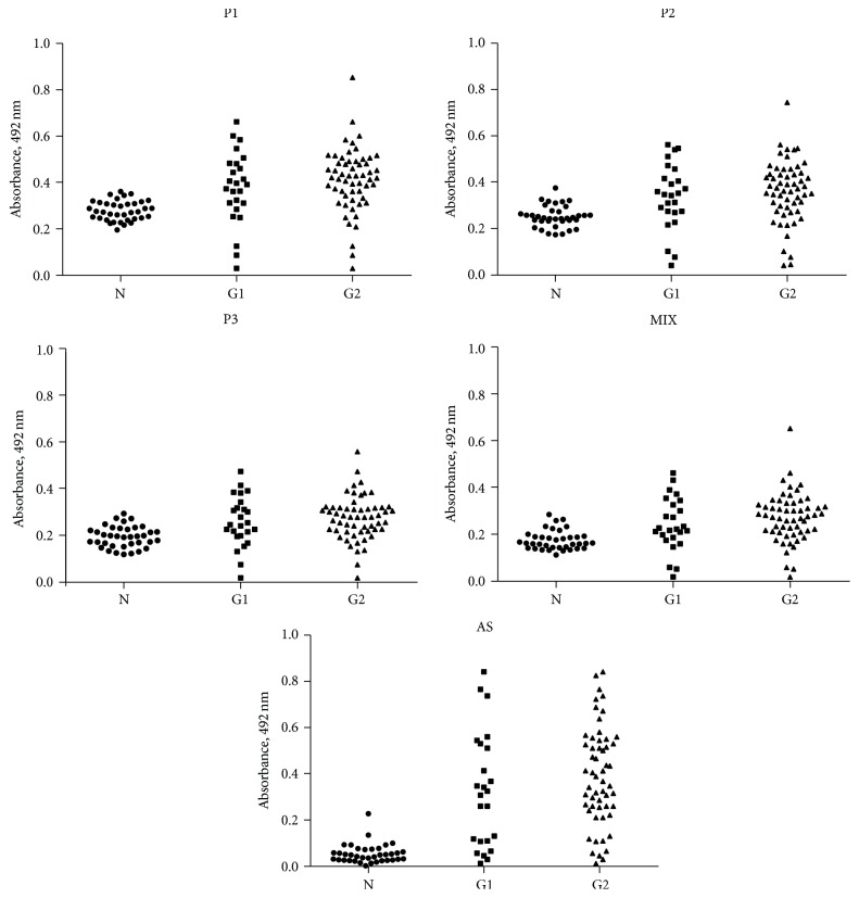
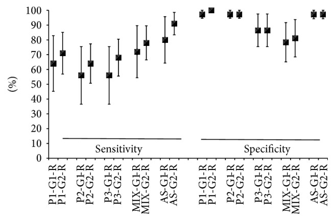
This work's goal was to research new candidate antigens for cutaneous leishmaniosis (CL). In order to reach the goal, we used random peptide phage display libraries screened using antibodies from Leishmania braziliensis patients. After selection, three peptides (P1, P2, and P3) were synthesized using Fmoc chemistry. The peptides individually or a mixture of them (MIX) was subsequently emulsified in complete and incomplete Freund's adjuvant and injected subcutaneously in golden hamsters. Sera from the hamsters administered with P1 presented antibodies that recognized proteins between 76 and 150 kDa from L. braziliensis. Sera from hamsters which had peptides P2 and P3, as well as the MIX, administered presented antibodies that recognized proteins between 52 and 76 kDa of L. braziliensis. The research on the similarity of the peptides' sequences in protein databases showed that they match a 63 kDa glycoprotein. The three peptides and the MIX were recognized by the sera from CL patients by immunoassay approach (ELISA). The peptides' MIX showed the best performance (79% sensitivity) followed by the P1 (72% sensitivity), and the AS presented 91% sensitivity. These results show a new route for discovering molecules for diagnosis or for immunoprotection against leishmaniosis.


Conflict of interest statement

The authors declare that they have no competing interests regarding the publication of this paper.

Figures

Figure 1
Figure 1
Reactivity of peptides with sera from immunized hamsters by ELISA. P1 peptide (a), P2 peptide (b), P3 peptide (c), and MIX of P1, P2, and P3 (d) were tested with anti-peptide serum P1 (P1), anti-peptide serum P2 (P2), anti-peptide serum P3 (P3), anti-peptide serum MIX (MIX), nonimmune serum, and serum against Freund's adjuvant. ELISA plates were coated with peptide (2 μg/mL) and incubated with hamster serum diluted at 1 : 50 (pool of sera with ten animals in each group). The detection reaction was conducted with anti-hamster IgG (whole molecule) peroxidase antibody (200 ng/mL).
Figure 2
Figure 2
Anti-peptide antibody reactivity to L. braziliensis antigens by western blot. Forty micrograms of L. braziliensis antigen was separated by 15% SDS-PAGE and then, using western blot, was reacted with anti-peptide serum P1 (lane 1), anti-peptide serum P2 (lane 2), anti-peptide serum P3, anti-peptide serum MIX, sera against Freund's adjuvant (lane 5), and NC = negative control → nonimmune serum (lane 6). Sera from the immunized hamster were tested at a 1 : 100 dilution. The detection reaction was performed with anti-hamster IgG (whole molecule) antibody conjugated to peroxidase (100 μg/mL).
Figure 3
Figure 3
Reactivity of peptides P1, P2, P3, MIX, and SA against sera from CL patients (G1: Group 1, G2: Group 2) and negative control (N).
Figure 4
Figure 4
Performance of ELISA test with synthetic peptides and sera of patients with cutaneous leishmaniosis. P1: peptide 1; P2: peptide 2; P3: peptide 3, MIX: P1 + P2 + P3; G1: Group 1 (patients with clinical cutaneous leishmaniosis and parasites isolated); G2: Group 2 (patients with clinical cutaneous leishmaniosis and serological diagnosis positive); SA: soluble antigen.

Similar articles

Cited by

References

    1. Desjeux P. Leishmaniasis: current situation and new perspectives. Comparative Immunology, Microbiology and Infectious Diseases. 2004;27(5):305–318. doi: 10.1016/j.cimid.2004.03.004. - DOI - PubMed
    1. Alvar J., Vélez I. D., Bern C., et al. Leishmaniasis worldwide and global estimates of its incidence. PLoS ONE. 2012;7(5) doi: 10.1371/journal.pone.0035671.e35671 - DOI - PMC - PubMed
    1. Szargiki R., de Castro E. A., Luz E., Kowalthuk W., Machado Â. M., Thomaz-Soccol V. Comparison of serological and parasitological methods for cutaneous leishmaniasis diagnosis in the state of Paraná, Brazil. Brazilian Journal of Infectious Diseases. 2009;13(1):47–52. doi: 10.1590/S1413-86702009000100011. - DOI - PubMed
    1. de Paiva-Cavalcanti M., de Morais R. C. S., Pessoa-e-Silva R., et al. Leishmaniases diagnosis: an update on the use of immunological and molecular tools. Cell and Bioscience. 2015;5(1, article 31) doi: 10.1186/s13578-015-0021-2. - DOI - PMC - PubMed
    1. Faber W. R., Oskam L., Van Gool T., et al. Value of diagnostic techniques for cutaneous leishmaniasis. Journal of the American Academy of Dermatology. 2003;49(1):70–74. doi: 10.1067/mjd.2003.492. - DOI - PubMed

MeSH terms

LinkOut - more resources