. 2017 Dec;55(1):1489-1499.
doi: 10.1080/13880209.2017.1307422.

Suppression of VEGF-induced angiogenesis and tumor growth by Eugenia jambolana, Musa paradisiaca, and Coccinia indica extracts

Affiliations

Suppression of VEGF-induced angiogenesis and tumor growth by Eugenia jambolana, Musa paradisiaca, and Coccinia indica extracts

Harsha Raj M et al. Pharm Biol. 2017 Dec.

Abstract

Context: Abnormal angiogenesis and evasion of apoptosis are hallmarks of cancer. Accordingly, anti-angiogenic and pro-apoptotic therapies are effective strategies for cancer treatment. Medicinal plants, namely, Eugenia jambolana Lam. (Myrtaceae), Musa paradisiaca L. (Musaceae), and Coccinia indica Wight & Arn. (Cucurbitaceae), have not been greatly investigated for their anticancer potential.

Objective: We investigated the anti-angiogenic and pro-apoptotic efficacy of ethyl acetate (EA) and n-butanol (NB) extracts of E. jambolana (seeds), EA extracts of M. paradisiaca (roots) and C. indica (leaves) with respect to mammary neoplasia.

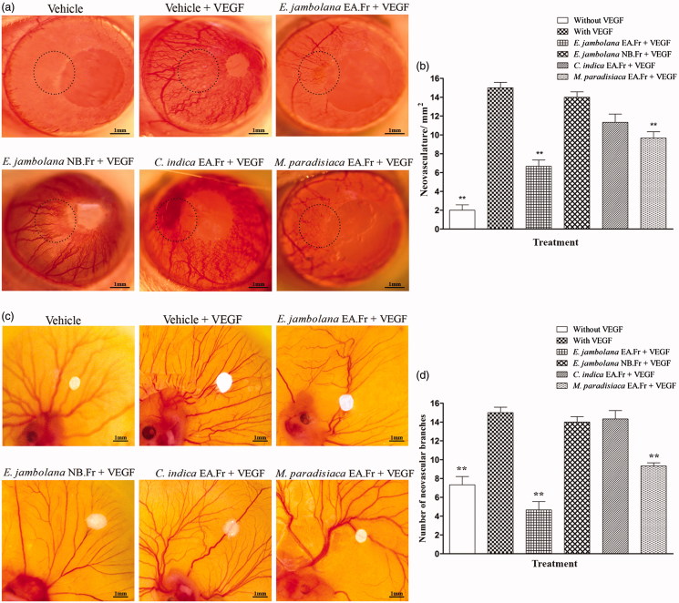
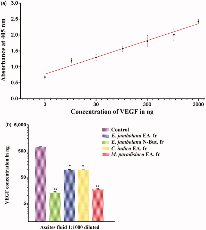
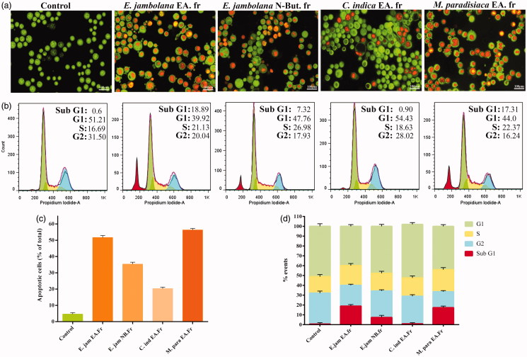
Materials and methods: Effect of extracts (2-200 μg/mL) on cytotoxicity and MCF-7, MDA-MB-231 and endothelial cell (EC) proliferation and in vitro angiogenesis were evaluated by MTT, 3[H]thymidine uptake and EC tube formation assays, respectively. In vivo tumour proliferation, VEGF secretion and angiogenesis were assessed using the Ehrlich ascites tumour (EAT) model followed by rat corneal micro-pocket and chicken chorioallantoic membrane (CAM) assays. Apoptosis induction was assessed by morphological and cell cycle analysis.

Results: EA extracts of E. jambolana and M. paradisiaca exhibited the highest cytotoxicity (IC50 25 and 60 μg/mL), inhibited cell proliferation (up to 81%), and tube formation (83% and 76%). In vivo treatment reduced body weight (50%); cell number (16.5- and 14.7-fold), secreted VEGF (∼90%), neoangiogenesis in rat cornea (2.5- and 1.5-fold) and CAM (3- and 1.6-fold) besides EAT cells accumulation in sub-G1 phase (20% and 18.38%), respectively.

Discussion and conclusion: Considering the potent anti-angiogenic and pro-apoptotic properties, lead molecules from EA extracts of E. jambolana and M. paradisiaca can be developed into anticancer drugs.

Keywords: Anti-angiogenic; cytotoxicity; pro-apoptotic.


Figures

Figure 1.
Figure 1.
Effects of E. jambolana, M. paradisiaca, and C. indica extracts on cell proliferation. (a) The metabolic and cytotoxic response of MDA-MB-231 cells assessed by MTT assay. MDA-MB-231 cells (3 × 104) were treated with plant extracts (2, 5, 10, 20, 40, 80, 100, and 200 μg/mL) for 24 h, washed with PBS, incubated for 4 h with MTT and the formazan crystals dissolved in DMSO and read at 570 nm. (b) Cell proliferation assessed by the rate of DNA synthesis using 3[H]thymidine. HUVEC, MCF-7 and MDA-MB-231 cells (3 × 104) were cultured in 12-well plates, serum starved and treated with plant extracts (20 μg/mL) along with rVEGF (10 ng) for 24 h. Post incubation for 4 h in presence of 3[H]thymidine, the DNA was harvested and rate of cell proliferation was measured in a liquid scintillation counter. (c) HUVEC tube formation assay. HUVECs (5 × 103) were cultured in EGM on Matrigel with plant extracts (20 μg/mL) along with rVEGF (10 ng), in a 96 well plate. After incubation for 24 h at 37 °C, capillary networks were photographed and quantified (Magnification: 200 ×). (d) Quantification of angiogenesis by counting the average number of branch points. Data in all results presented as mean ± SEM of three independent experiments; **p < 0.01 and *p < 0.05.
Figure 2.
Figure 2.
Effects of plant extracts on tumour growth and angiogenesis in vivo. (a) Body weights of EAT-bearing; untreated and treated with plant extracts were recorded. 6th day onwards, plant extracts (100 mg/kg body weight) was administered (i.p) for six days. The animals were sacrificed on the 12th day. EAT cells were collected along with ascites fluid and centrifuged, (b) secreted ascited volume, (c) EAT cell count assessed by trypan blue dye exclusion method, (d) representative photographs of the peritoneum of the untreated and treated mice and (e) peritoneal MVD marked by arrows in each peritoneum assessed by H and E staining of sections. At least five mice were used in each group and the results obtained are an average of three individual experiments and means of ± S.E.M. n = 5 per group.
Figure 3.
Figure 3.
Effects of plant extracts on neovasculature in the rat cornea and chick embryo CAM assays. (a) Representative photographs of VEGF-induced rat corneal neovascularization, plant extracts 20 μg/hydron polymer pellet were surgically implanted into the micro-pocket in the cornea of one eye. On day 7, the extent of neovascularization or inhibition was visualized and photographed under a dissection microscope. (b) Quantitative comparison of the number of neovascular vessels per mm2 was estimated. (c) Representative photographs of VEGF-induced neovascularization in shell-less CAM of chicken embryos. Plant extracts, 20 μg/filter disc was placed on the CAM of 6-day old chicken embryos. After 72 h of incubation, the area surrounding the filter disc was inspected for changes in neovascularization. (d) Quantitative comparison of the number of neovascular branches surrounding the plant extract containing filter paper discs. The data shown represent the results of experiments that were performed using a maximum of 5 eggs in each group. All quantitative data are presented as mean ± SEM of five independent counts; **p < 0.01.
Figure 4.
Figure 4.
Effects of plant extracts on the secretion of VEGF in ascites fluid of tumour bearing mouse. Indirect ELISA was carried out using the ascites fluid harvested from the control (tumour bearing) and plant extracts treated (100 mg/kg body weight) mice to quantify the VEGF in ascites fluid using anti-VEGF165 antibodies. (a) ELISA standard curve for VEGF. (b) The histogram showing a comparison of VEGF levels in the ascites of untreated control and treated groups. Data are representative of three independent experiments and values are expressed in mean ± SEM, **p < 0.01 and *p < 0.05.
Figure 5.
Figure 5.
Apoptosis induction and cell cycle analysis of plant extract treated EAT cells. (a) In vivo treated EAT cells with or without plant extracts were stained with acridine orange and ethidium bromide and verified for apoptotic characteristics such as plasma membrane blebbing, chromatin condensation and apoptotic body formation under a fluorescence microscope. (b) Effect of plant extracts on cell cycle progression. EAT cells, in vitro, were treated with or without different plant extracts (20 μg/mL). After 24 h of treatment, distribution of cell cycle phases and apoptotic cell population was quantitated based on flow cytometric analysis. (c) A quantitative comparison of the number of apoptotic cells in 10 random fields. (d) Bar diagram showing the percentage of cells present in different phases of cell cycle.

Similar articles

Cited by

References

    1. Afsar T, Trembley JH, Salomon CE, Razak S, Khan MR, Ahmed K.. 2016. Growth inhibition and apoptosis in cancer cells induced by polyphenolic compounds of Acacia hydaspica: involvement of multiple signal transduction pathways. Sci Rep. 6:23077. - PMC - PubMed
    1. Ali EM, Sheta M, El Mohsen MA.. 2011. Elevated serum and tissue VEGF associated with poor outcome in breast cancer patients. Alexandria J Med. 47:217–224.
    1. Andzi-Barhé T, Massala KK, Obame LC, Engonga JL.. 2015. Phytochemical studies, total phenolic and flavonoids content and evaluation of antiradical activity of the extracts of the leaves from Dischistocalyx sp. (Acanthaceae). J Pharmacogn Phytochem. 3:174–178.
    1. Baliga MS.2011. Anticancer, chemopreventive and radioprotective potential of black plum (Eugenia jambolana Lam.). Asian Pac J Cancer Prev. 12:3–15. - PubMed
    1. Bikfalvi A, Bicknell R.. 2002. Recent advances in angiogenesis, anti-angiogenesis and vascular targeting. Trends Pharmacol Sci. 23:576–582. - PubMed

MeSH terms

LinkOut - more resources