. 2017 Apr 3:8:14967.
doi: 10.1038/ncomms14967.

A brain-sparing diphtheria toxin for chemical genetic ablation of peripheral cell lineages

Affiliations

A brain-sparing diphtheria toxin for chemical genetic ablation of peripheral cell lineages

Mafalda M A Pereira et al. Nat Commun. .

Erratum in

Abstract

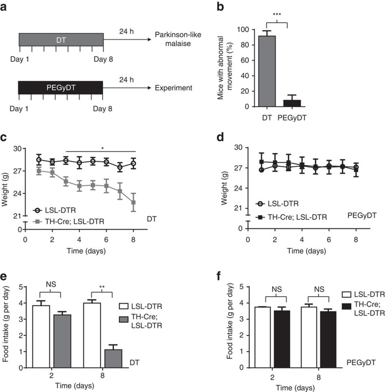
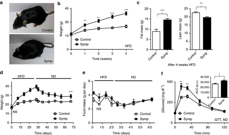
Conditional expression of diphtheria toxin receptor (DTR) is widely used for tissue-specific ablation of cells. However, diphtheria toxin (DT) crosses the blood-brain barrier, which limits its utility for ablating peripheral cells using Cre drivers that are also expressed in the central nervous system (CNS). Here we report the development of a brain-sparing DT, termed BRAINSPAReDT, for tissue-specific genetic ablation of cells outside the CNS. We prevent blood-brain barrier passage of DT through PEGylation, which polarizes the molecule and increases its size. We validate BRAINSPAReDT with regional genetic sympathectomy: BRAINSPAReDT ablates peripheral but not central catecholaminergic neurons, thus avoiding the Parkinson-like phenotype associated with full dopaminergic depletion. Regional sympathectomy compromises adipose tissue thermogenesis, and renders mice susceptible to obesity. We provide a proof of principle that BRAINSPAReDT can be used for Cre/DTR tissue-specific ablation outside the brain using CNS drivers, while consolidating the link between adiposity and the sympathetic nervous system.


Conflict of interest statement

The authors declare no competing financial interests.

Figures

Figure 1
Figure 1. PEGylation of DT and its in vitro functionality after modification.
(a) PEG residues bind to the lysines in DT. (b) Schematic representation of PEGylation of DT through conjugation of NHS-PEG4 at lysines. (c) Mass spectrometry of DT (left) and PEGyDT (right) samples. DT peak is identified in red and PEGyDT peaks are identified in green. (d) Live- and dead-cell populations after 48 h of incubation with vehicle, DT and PEGyDT in HeLa cells. (e) Time course of cell death after incubation with vehicle, DT and PEGyDT (***P<0.0001, n=3). Statistics were performed using one-way ANOVA test followed by Tukey test. Data are represented as mean±s.e.m. (related to Supplementary Figs 1 and 2).
Figure 2
Figure 2. BRAINSPAReDT ablates peripheral neurons but not those in the brain.
(a) Confocal microscopy imaging of TH+ neurons after i.p. injection of vehicle, DT or PEGyDT. Scale bar, 250 μm. (b) Confocal microscopy imaging of nerve fibres in fat stained for TH and for β3-Tub after i.p. injection of vehicle, DT or PEGyDT. Scale bar, 100 μm. (c) Quantification of TH+/DAPI neurons after i.p. injection of vehicle, DT or PEGyDT (***P<0.0001, n=3). (d) Quantification of TH+3-Tub neurons after in vivo injection of vehicle, DT or PEGyDT (***P<0.0001, n=3). Statistics were performed using one-way ANOVA test followed by Tukey test. Data are represented as mean±s.e.m. (related to Supplementary Figs 3 and 4a). NS, not significant.
Figure 3
Figure 3. BRAINSPAReDT prevents Parkinson-like movements and decaying health.
(a) Schematic representation of the experimental design. (b) Scoring for abnormal movement of DT and PEGyDT-injected mice (***P<0.0001, n=4). (c) Mice weight during daily administration of 0.02 pmol g−1 of body weight of DT (*P<0.01, n=4). (d) Mice weight during daily administration of 0.02 pmol g−1 of body weight of PEGyDT (n=5). (e) Food intake on the first and last day of administration of 0.02 pmol g−1 of body weight of DT (**P<0.001, n=3). (f) Food intake on the first and last day of administration of 0.02 pmol g−1 of body weight of PEGyDT (n=3). Statistics were performed using unpaired t-test. Data are represented as mean±s.e.m. NS, not significant.
Figure 4
Figure 4. Sympathectomy with BRAINSPAReDT impairs adaptive thermogenesis and NE content in adipose tissues.
(a) Body temperature during cold (4 °C) exposure (***P<0.0001, n=5). Controls are PEGyDT-injected LSL-DTR mice, which are hereafter referred to as ‘Control'. (b) Norepinephrine content of SubQ adipose tissue and BAT after 8 h of cold exposure (****P<0.00001, n=4 for Symp and n=6 for Control). (c) mRNA levels of browning genes in SubQ adipose tissue after 8 h of cold exposure (#P=0.066, **P<0.001, ****P<0.00001, n=9). (d) Body weight change and food intake during a leptin challenge (*P<0.01, n=4). Statistics were performed using unpaired t-test. Data are represented as mean±s.e.m. (related to Supplementary Fig. 4b,c). NS, not significant.
Figure 5
Figure 5. Sympathectomy with BRAINSPAReDT predisposes mice to sustained obesity and glucose intolerance without affecting food intake.
(a) TH-Cre; LSL-DTR and control LSL-DTR mice after 4 weeks of HFD regimen. (b) Weight gain during 4 weeks on HFD, after ablation (**P<0.001, ***P<0.0001, n=6). (c) Nuclear magnetic resonance analysis of fat and lean mass (***P<0.0001, **P<0.001, n=8). (d) Weight variation before and after HFD withdrawal (***P<0.0001, n=6). (e) Food intake before and after HFD withdrawal. (f) GTT during ND (*P<0.01, **P<0.001, n=8). Statistics were performed using unpaired t-test. Data are represented as mean±s.e.m. (related to Supplementary Fig. 5). GTT, glucose tolerance test.
Figure 6
Figure 6. Obese mice sympathectomized with BRAINSPAReDT have deficits in thermogenic gene expression and nocturnal hypoactivity but normal energy expenditure.
(a) Thermogenic gene expression in SubQ adipose tissue (*P<0.01, **P<0.001, ***P<0.0001, n=6). (b) Thermogenic gene expression in BAT (*P<0.01, n=7 for Symp and n=−8 for Control). The a.u. used in a,b is the same. The data were normalized to GAPDH and SYBR Green was used as a probe. (c) Locomotor activity, energy expenditure and respiratory quotient (**P<0.001, n=8). Statistics were performed using unpaired t-test. Data are represented as mean±s.e.m. (related to Supplementary Figs 6 and 7). EE, energy expenditure; NS, not significant; RQ, respiratory quotient.

References

    1. Picklo M. J. Methods of sympathetic degeneration and alteration. J. Auton. Nerv. Syst. 62, 111–125 (1997). - PubMed
    1. Beznák A. B. L. & Hasch Z. The effect of sympathectomy on the fatty deposit in connective tissue. Q. J. Exp. Physiol. 27, 5–12 (1937).
    1. Cousin B. et al.. Local sympathetic denervation of white adipose tissue in rats induces preadipocyte proliferation without noticeable changes in metabolism. Endocrinology 133, 2255–2262 (1993). - PubMed
    1. Youngstrom T. G. & Bartness T. J. White adipose tissue sympathetic nervous system denervation increases fat pad mass and fat cell number. Am. J. Physiol. 275, 1488–1493 (1998). - PubMed
    1. Zeng W. et al.. Sympathetic neuro-adipose connections mediate leptin-driven lipolysis. Cell 163, 84–94 (2015). - PMC - PubMed

Publication types

MeSH terms