. 2017 Apr 3:8:14932.
doi: 10.1038/ncomms14932.

Macrocycle peptides delineate locked-open inhibition mechanism for microorganism phosphoglycerate mutases

Affiliations

Macrocycle peptides delineate locked-open inhibition mechanism for microorganism phosphoglycerate mutases

Hao Yu et al. Nat Commun. .

Abstract

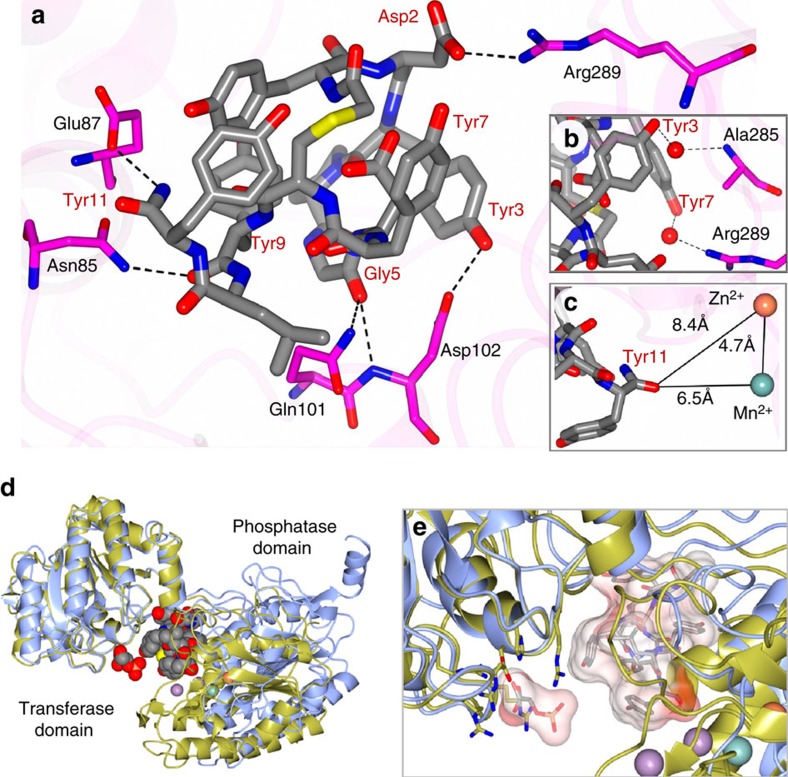
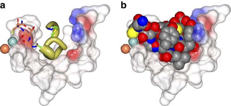
Glycolytic interconversion of phosphoglycerate isomers is catalysed in numerous pathogenic microorganisms by a cofactor-independent mutase (iPGM) structurally distinct from the mammalian cofactor-dependent (dPGM) isozyme. The iPGM active site dynamically assembles through substrate-triggered movement of phosphatase and transferase domains creating a solvent inaccessible cavity. Here we identify alternate ligand binding regions using nematode iPGM to select and enrich lariat-like ligands from an mRNA-display macrocyclic peptide library containing >1012 members. Functional analysis of the ligands, named ipglycermides, demonstrates sub-nanomolar inhibition of iPGM with complete selectivity over dPGM. The crystal structure of an iPGM macrocyclic peptide complex illuminated an allosteric, locked-open inhibition mechanism placing the cyclic peptide at the bi-domain interface. This binding mode aligns the pendant lariat cysteine thiolate for coordination with the iPGM transition metal ion cluster. The extended charged, hydrophilic binding surface interaction rationalizes the persistent challenges these enzymes have presented to small-molecule screening efforts highlighting the important roles of macrocyclic peptides in expanding chemical diversity for ligand discovery.


Conflict of interest statement

The authors declare no competing financial interests.

Figures

Figure 1
Figure 1. Species-dependent phosphoglycerate mutase catalytic mechanisms and overview of affinity selection.
(a) Isomerization catalysed by PGMs illustrating the phosphohistidine enzyme/2,3-phosphoglycerate intermediate of human cofactor-dependent PGM (top) and the phosphoserine enzyme intermediate of C. elegans cofactor-independent PGM. (b) Random nonstandard peptide integrated discovery (RaPID) begins with an mRNA library encoding trillions of potential peptides 6–14 amino acids in length. The mRNA library is ligated to an adapter incorporating the amino nucleoside, puromycin. The flexible in vitro translation (FIT) system is used to create the peptide library with an L- or D-N-chloroacetyl tyrosine (dark blue sphere) charged initiator tRNA and 19 proteogenic amino acids (grey spheres), methionine is excluded as its tRNA is charged with the chloroacetyl tyrosine. Incorporation of a cysteine during translation results in macrocyclization via thiolate nucleophilic attack on the chloroacetyl electrophile. After incubating with the library the beads are washed to enrich the bound conjugates which are then reverse transcribed and amplified via PCR. PCR products are transcribed to mRNAs and the process is repeated or PCR products sequenced to reveal the peptide sequences captured. Peptides from these sequences are then produced in milligram quantity using solid-phase peptide synthesis (SPPS).
Figure 2
Figure 2. Pharmacologic–phylogenetic relationship of iPGM macrocyclic peptide inhibitor.
(a) Structure of the Ce-2 macrocycle obtained from affinity selection showing truncation to give Ce-2d and position of Cys14Ser substitution. Note the thioether bond (yellow), D-tyrosine (blue), and thiol of Cys14 (light red). (b) IC50 dependence of Ce-2 on C. elegans iPGM enzyme concentration (50 pM to 1 μM), grey region indicated concentration range for PGMs used in initial inhibitory activity profiling obtained in Table 1. (c) Concentration-response curves for characterization of Ce-2d on the iPGM orthologues and dPGM isozymes. (d) Phylogenetic tree constructed for amino-acid sequence alignments of seven species orthologues and isozymes of PGM. Percentage bootstrap values based on 1,000 replicates are indicated at branch nodes. (e) Concentration-response curves for characterization of Ce-2S on the iPGM orthologues and dPGM isozymes. All data determined from the enzyme-coupled bioluminescent assay; PGM concentrations as indicated in Table 1. Plots are representatives from individual experiments (N=3); error bars are standard deviations values of technical replicates.
Figure 3
Figure 3. Ce-2d traps iPGM in an open conformation.
(a) One subunit of the asymmetric unit showing the binding mode of the Ce-2d macrocycle to C. elegans iPGM. The Mn2+ and Zn2+ ions are represented as blue and tan spheres, respectively, and the bound macrocycle is drawn as CPK space-filling spheres in a cavity defined by iPGM residues within 5 Å (transparent spheres). (b) Superposition of C. elegans iPGM-o (cyan), C. elegans iPGM-m (tan) and C. elegans iPGM·Ce-2d (aquamarine). The Ce-2d peptide is represented as cylinders. (c) Macrocycle (cylinders) positioned within a cleft of iPGM represented as an electrostatic surface. (d) CPK space-filling representations of Ce-2d illustrating the ‘capping' orientation of the five tyrosine residues (1, 3, 7, 9 and 11) and (e) the edge-to-face interaction of Tyr 1 and 9. Additional residues are indicated.
Figure 4
Figure 4. Ce-2d · iPGM interactions.
(a) Hydrogen bond interactions (black dashed lines) between C. elegans iPGM and Ce-2d. Direct interactions and (b) water (red spheres) mediated contacts. (c) Distance from the C-terminal amide of Tyr11 and the Zn2+ and Mn2+ ion centres. (d) Superimposed structure of C. elegans iPGM·Ce-2d with that of Staphylococcus aureus iPGM in 2-phosphoglyceric acid bound form (PDB: 4NWX). The following, S. Aureus : C. elegans residue pairs were used for alignment: 123His147, 153-154Asp177-178, 191Arg216, 185Arg210, 257Arg284 and 260Arg287. Ce-2d and 2-PG are shown as CPK space filling models. The purple spheres are the Mn2+ ions of S. aureus iPGM and the blue and tan spheres are the Mn2+ and Zn2+ ion, respectively of C. elegans iPGM. (e) Enlarged region from d showing the relative locations of the 2-PG and Ce-2d as cylinder models with transparent van der Waals surfaces and alignment residue side chains clustering around 2-PG.
Figure 5
Figure 5. Structural basis underlying the pharmacologic–phylogenetic Ce-2 macrocycle series-iPGM orthologue relationship.
(a) Relationship between Ce-2 macrocycle truncation series IC50s and iPGM orthologues. Analogues with no detectable inhibitory activity are indicated as inactive. Data represent mean±s.d. values of the log normal distributed IC50s determined for the given peptide for experiments with ≥4 replicates; otherwise error bar is determined from the nonlinear fit of the standard Hill equation to the aggregated data from the replicates. Values are from Supplementary Table 3 converted from pIC50 where IC50=10-pIC50. (b) Select amino-acid sequence alignment of iPGM orthologues in this study (see Supplementary Fig. 10 for full alignment). iPGM residues within 5 Å of Ce-2d are coloured orange. Residues identical between C. elegans and E. coli iPGM are coloured yellow; grey indicated hinge regions; green and blue are amino acids that ligand metal ions. (c) Cavity formed from C. elegans iPGM residues (light blue chain under transparent spheres) within 5 Å of the Ce-2d macrocycle shown as a worm α-chain (gold) representation scaled by B-factor with select side chains (Tyr3, Pro4, thioether linkage, and C-terminal Tyr11 amide) shown. The iPGM Ala334 residue is shown as a CPK space fill. Electrostatic surface of the Ce-2d binding cavity is also shown.
Figure 6
Figure 6. Modelling of C-terminal residues of Ce-2 onto Ce-2d.
(a) The Ce-2d macrocycle is shown as worm α-chain (gold) representation scaled by B-factor within a cavity of C. elegans iPGM residues (transparent spheres) formed from residues within 5Å of cyclic peptide. The C-terminal residues, -Gly12-Thr13-Cys14-Gly15 of Ce-2 were modelled onto the iPGM·Ce-2d complex and are shown as tan sticks extending from Ce-2d. Electrostatic surface of the binding cavity is also shown. (b) Ce-2 van der Waals radii shown using a CPK model. The Cys14 sulfhydryl is shown in yellow.

Similar articles

Cited by

References

    1. Coghlan A. Nematode Genome Evolution ed. Community, T.C.e.R. WormBook (2005). - PMC - PubMed
    1. World Health Organization. The World Health Report: 2004: Changing History (2004).
    1. Churcher T. S. et al.. Identifying sub-optimal responses to ivermectin in the treatment of River Blindness. Proc. Natl Acad. Sci. USA 106, 16716–16721 (2009). - PMC - PubMed
    1. Osei-Atweneboana M. Y. et al.. Phenotypic evidence of emerging ivermectin resistance in Onchocerca volvulus. PLoS Negl. Trop. Dis. 5, e998 (2011). - PMC - PubMed
    1. Kumar S. et al.. Mining predicted essential genes of Brugia malayi for nematode drug targets. PLoS ONE 2, e1189 (2007). - PMC - PubMed

Publication types

MeSH terms