Review
. 2017 May;17(5):281-294.
doi: 10.1038/nri.2017.19. Epub 2017 Apr 3.

Mechanisms of central tolerance for B cells

Affiliations
Review

Mechanisms of central tolerance for B cells

David Nemazee. Nat Rev Immunol. 2017 May.

Abstract

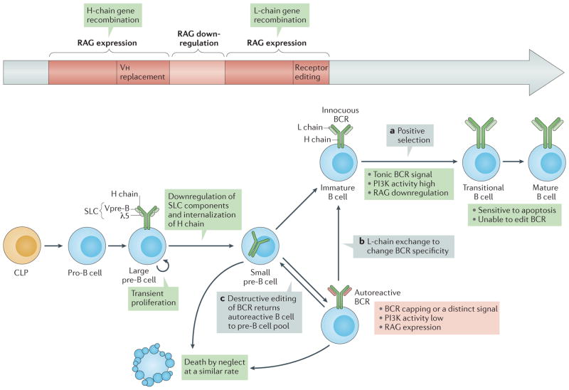
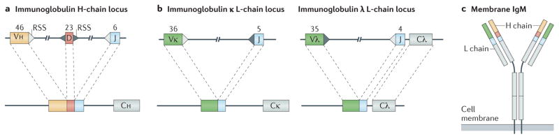
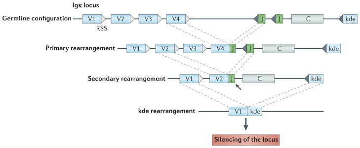
Immune tolerance hinders the potentially destructive responses of lymphocytes to host tissues. Tolerance is regulated at the stage of immature B cell development (central tolerance) by clonal deletion, involving apoptosis, and by receptor editing, which reprogrammes the specificity of B cells through secondary recombination of antibody genes. Recent mechanistic studies have begun to elucidate how these divergent mechanisms are controlled. Single-cell antibody cloning has revealed defects of B cell central tolerance in human autoimmune diseases and in several human immunodeficiency diseases caused by single gene mutations, which indicates the relevance of B cell tolerance to disease and suggests possible genetic pathways that regulate tolerance.


Conflict of interest statement

Competing interests statement

The author declares no competing interests.

Figures

Figure 1
Figure 1. B cell development
Pro-B cells in the bone marrow, which are derived from common lymphoid progenitor (CLP) cells, initiate heavy (H)-chain gene rearrangement through expression of recombination-activating genes (RAG1 and RAG2; collectively referred to here as RAG) and epigenetic modifications of the H-chain loci that promote accessibility. Productive H-chain gene assembly leads to the association of IgM H-chain (μ-chain) protein with surrogate light-chain (SLC) components λ5 (also known as IGLL1) and Vpre-B, and surface expression of the pre-B cell receptor (pre-BCR) in large pre-B cells,. Pro-B cells can also undergo H-chain variable region (Vh) replacement reactions, whereby Vh elements recombine to a conserved heptamer within the Vh element of an already rearranged VDJ exon. Spontaneous, antigen-independent triggering of the pre-BCR promotes progression to the large pre-B cell stage, which involves downregulation of RAG expression and transient proliferation. Differentiation to the small pre-B cell stage follows; at this stage, SLC components are downregulated, RAG is re-expressed and RAG activity is redirected to the L-chain genes. L chains that pair with H chains trigger tonic BCR signalling, which promotes positive selection when the BCR is non-autoreactive (part a) or receptor editing (parts b and c) when the BCR is autoreactive or if tonic signalling is impaired. Editing can lead to exchange of one functional L chain for another, which can render the BCR innocuous and allow developmental progression (part b), or to secondary rearrangements that prevent L-chain expression (part c), such as out-of-frame joins that destroy the original L-chain gene but fail to replace it, which returns the cell to the pre-B cell compartment,. Cells that go through positive selection enter the transitional B cell stage, at which stage B cells seem to be extremely sensitive to apoptosis while losing the ability to edit the BCR,. Small pre-B cells and editing B cells have a similar turnover rate, which I interpret to indicate that they die ‘by neglect’ owing to prolonged insufficiency of phosphoinositide 3-kinase (PI3K)–AKT activity.
Figure 2
Figure 2. Antibody gene assembly by DNA recombination between gene segments
The variable part of antibody genes is composed of variable (V), diversity (D) and joining (J) elements on the locus encoding the heavy (H) chain (part a), and V and J elements on each of two loci encoding L chains, λ and κ (part b). Triangles show the recombination signal sequences (RSSs) adjacent to the gene segments. Numbers above each element indicate the estimated sums of the indicated gene elements arranged along the human immunoglobulin loci. In addition to the coding element shown, each V region has its own upstream promoter, leader exon and intron (not shown). Each V, D and J element can recombine as shown to generate many combinations of elements within a locus. On the H-chain locus, D-to-J rearrangements occur first, followed by V-to-DJ recombination. The cartoon of membrane IgM protein (part c) shows the approximate placement of amino acid residues encoded by each element. Also shown is the transmembrane region of IgM and the short cytoplasmic tail, which has the sequence Lys-Val-Lys. C, constant region.
Figure 3
Figure 3. B cell receptor signalling
In the assembled B cell receptor (BCR), CD79A–CD79B is weakly bound by Src family kinases (SFKs). Antigen binding promotes tyrosine phosphorylation of CD79A and CD79B on their immunoreceptor tyrosine-based activation motifs (ITAMs),. Phosphorylated ITAMs recruit spleen tyrosine kinase (SYK) and upregulate its kinase activity,. CD19 functions as a BCR co-receptor, leading to tyrosine phosphorylation within YXXM motifs in the cytoplasmic tail of CD19 that recruit the p85 regulatory subunit of phosphoinositide 3-kinase (PI3K),, (left). PI3K activation mediated through this pathway or by the adaptor protein B cell adaptor for PI3K (BCAP) mediates phosphorylation of phosphatidylinositol-4,5-bisphosphate (PtdInsP2), generating phosphatidylinositol-3,4,5-trisphosphate (PtdInsP3), which recruits AKT, 3-phosphoinositide-dependent protein kinase 1 (PDK1), Bruton’s tyrosine kinase (BTK) and other enzymes that are essential for signal propagation. In a distinct pathway (right), B cell linker protein (BLNK) functions as a scaffold and substrate for SYK- and SFK-mediated phosphorylation, promoting the recruitment of phospholipase Cγ2 (PLCγ2), BTK, VAV guanine nucleotide exchange factor proteins and growth factor receptor-bound protein 2 (GRB2). Activated PLCγ2 hydrolyses PtdInsP2 to diacylglycerol (DAG) and inositol-1,4,5-trisphosphate (InsP3), which promotes Ca2+ mobilization through the InsP3 receptor (InsP3R) and opening of the plasma membrane Ca2+ channel ORAI. DAG recruits protein kinase C (PKC) isoforms and RAS guanyl-releasing proteins (RASGRPs). During positive selection of immature B cells, AKT activity suppresses forkhead box protein O1 (FOXO1) nuclear localization, which turns off recombination-activating gene (RAG) expression, and increases production of the transcription factor MYC, which (together with AKT) promotes cell survival. Another BCR-triggered pathway involves the guanine nucleotide exchange factor son of sevenless 1 (SOS1), the small GTPase RAS, the serine kinase RAF1, MAPK/ERK kinase (MEK) and extracellular signal-regulated kinases (ERKs). Ca2+ mobilization through InsP3R and ORAI promotes the activation of calcineurin and the nuclear localization of nuclear factor of activated T cells (NFAT). PKC activation promotes activation of nuclear factor−κB (NF−κB). PI3K and its downstream activities seem to be promoted in immature B cells by the unligated BCR, whereas BCR ligation activates the BLNK pathway transiently. This leads to BCR internalization and reduced signalling through both pathways, and drives RAG expression, developmental arrest and cell starvation. AP-1, activator protein 1; ER, endoplasmic reticulum; PTEN, phosphatase and tensin homologue; STIM1, stromal interaction molecule 1. Adapted with permission from REF. , F1000Research.
Figure 4
Figure 4. Recombination events associated with receptor editing on the immunoglobulin κ locus can silence or alter gene expression
The top line of the figure shows the germline configuration of gene segments in the human immunoglobulin κ (Igκ) locus, with variable (V) and joining (J) elements flanked by recombination signal sequences (RSSs). Also shown is the κ deleting element (kde). The first rearrangement on a locus is known as the primary rearrangement. Secondary rearrangement can silence the primary rearrangement; for example, in this case, V2 rearrangement to a downstream J element replaces the V4–J join. Note that some Vκ genes rearrange by inversion rather than excision of intervening DNA (not shown). When they are in-frame, secondary rearrangements can replace one functional V region with another. However, like all immunoglobulin gene rearrangements, these rearrangements are often non-functional and so can silence κ light-chain protein expression rather than replace it. Out-of-frame primary rearrangements can similarly be displaced by secondary rearrangements. Even loci that have ‘used up’ the J elements (arrow) can silence the locus by subsequent kde recombination (bottom line). The kde has no coding function, but behaves as a non-functional, but rearrangeable, J element, leading to deletion of the constant (C) region and silencing of the locus. Such rearrangements are often found in cells expressing λ light chains. The λ locus in humans has a similar organization, although a kde-like element has not been identified.

References

    1. Duong BH, et al. Decoration of T-independent antigen with ligands for CD22 and Siglec-G can suppress immunity and induce B cell tolerance in vivo. J Exp Med. 2010;207:173–174. - PMC - PubMed
    1. Macauley MS, et al. Antigenic liposomes displaying CD22 ligands induce antigen-specific B cell apoptosis. J Clin Invest. 2013;123:3074–3083. - PMC - PubMed
    1. Sabouri Z, et al. Redemption of autoantibodies on anergic B cells by variable-region glycosylation and mutation away from self-reactivity. Proc Natl Acad Sci USA. 2014;111:E2567–E2575. - PMC - PubMed
    1. Goodnow CC, et al. Altered immunoglobulin expression and functional silencing of self-reactive B lymphocytes in transgenic mice. Nature. 1988;334:676–682. A monumental paper that uses immunoglobulin transgenic mice to show that clonal anergy is a mechanism of B cell tolerance. - PubMed
    1. Klinman NR. The “clonal selection hypothesis” and current concepts of B cell tolerance. Immunity. 1996;5:189–195. - PubMed

LinkOut - more resources