. 2017 Apr 1;9(4):68.
doi: 10.3390/v9040068.

ICAM-1 Binding Rhinoviruses Enter HeLa Cells via Multiple Pathways and Travel to Distinct Intracellular Compartments for Uncoating

Affiliations

ICAM-1 Binding Rhinoviruses Enter HeLa Cells via Multiple Pathways and Travel to Distinct Intracellular Compartments for Uncoating

Haleh Ganjian et al. Viruses. .

Abstract

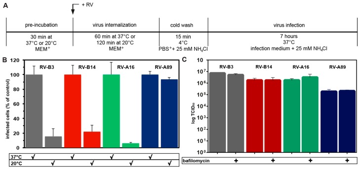
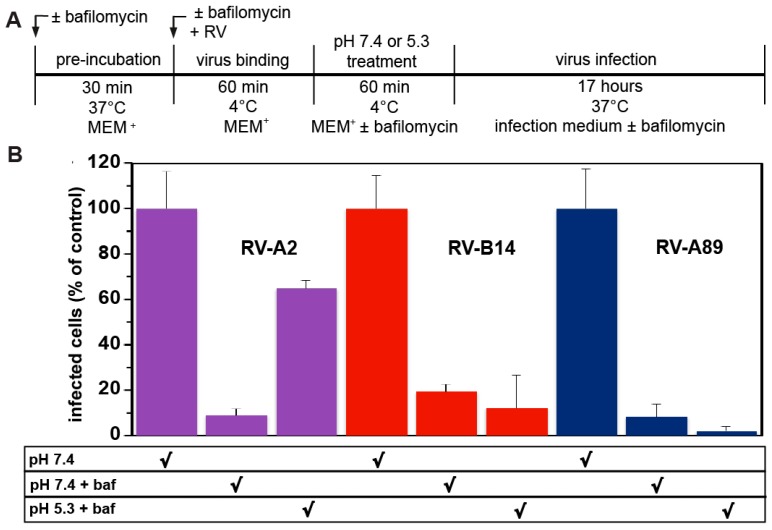
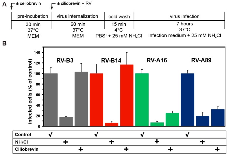
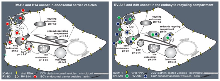
Of the more than 150 human rhinovirus (RV) serotypes, 89 utilize intercellular adhesion molecule-1 (ICAM-1) for cell entry. These belong either to species A or B. We recently demonstrated that RV-B14 and RV-A89, despite binding this same receptor, are routed into distinct endosomal compartments for release of their RNA into the cytosol. To gain insight into the underlying mechanism we now comparatively investigate the port of entry, temperature-dependence of uncoating, and intracellular routing of RV-B3, RV-B14, RV-A16, and RV-A89 in HeLa cells. The effect of various drugs blocking distinct stages on the individual pathways was determined via comparing the number of infected cells in a TissueFaxs instrument. We found that RV-B14 and RV-A89 enter via clathrin-, dynamin-, and cholesterol-dependent pathways, as well as by macropinocytosis. Drugs interfering with actin function similarly blocked entry of all four viruses, indicating their dependence on a dynamic actin network. However, uniquely, RV-A89 was able to produce progeny when internalized at 20 °C followed by neutralizing the endosomal pH and further incubation at 37 °C. Blocking dynein-dependent endosomal transport prevented uncoating of RV-A16 and RV-A89, but not of RV-B3 and RV-B14, indicative for routing of RV-A16 and RV-A89 into the endocytic recycling compartment for uncoating. Our results call for caution when developing drugs aimed at targeting entry or intracellular trafficking of all rhinovirus serotypes.

Keywords: ICAM-1; actin; clathrin; dynamin; dynein; endocytosis; human rhinoviruses; macropinocytosis; productive uncoating.


Conflict of interest statement

The authors declare no conflict of interest. The founding sponsors had no role in the design of the study; in the collection, analyses, or interpretation of data; in the writing of the manuscript, and in the decision to publish the results.

Figures

Figure 1
Figure 1
RV-B14 and RV-A89 enter HeLa cells via multiple pathways. The experimental setup is depicted in (A). After pre-incubating cells ± drugs (NH4Cl, dynasore, chlorpromazine, filipin, or blebbistatin), the respective virus at 100 TCID50/cell (50% tissue culture infectious dose/cell) was internalized for 60 min ± drugs. Non-internalized viruses were washed off with ice-cold phosphate buffered saline containing 1 mM CaCl2 and 1 mM MgCl2 (PBS+) plus NH4Cl to raise the endosomal pH and thereby stop further virus uncoating. Cells were transferred into infection medium containing 25 mM NH4Cl and dsRNA stemming from replicating viruses was detected with mAbJ2, followed by Alexa-488 goat-anti mouse IgG. Nuclei were stained with 4′,6-diamidino-2-phenylindol DAPI. The number of cells revealing mAbJ2 staining was determined in a TissueFaxs and related to the total number of cells. Infected cells in the absence of any drug were set to 100%. In each sample about 500,000 cells were analysed. (B) Data shown are the mean (± standard deviation (SD)) of five separate experiments each carried out with five parallels. The presence of NH4Cl throughout the incubation served as negative control. In all experiments shown (also in Figure 2, Figure 3, Figure 4 and Figure 5), NH4Cl blocked viral uncoating by 80–90%. Based on the inhibitory effect of dynasore, chlorpromazine, filipin, and blebbistatin, RV-B14 and RV-A89 enter HeLa cells by dynamin, clathrin, and cholesterol-dependent pathways, as well as by macropinocytosis. (C) HeLa cells were transfected with plasmids encoding myc-tagged dominant-negative mutants of AP-180 (AP180-C) and amphiphysin (amp-SH3), as indicated. After 24 h, RV-B14 or RV-A89 (100 TCID50/cell) was internalized for 60 min at 37 °C, cells were cooled, viruses in the supernatant medium were removed by washing, and myc-tagged proteins were stained with anti-myc antibodies (green) and viral proteins with rat anti-RV-14 antibody or antiserum P5, followed by the respective secondary antibodies (red). Bar, 10 µm. Cells expressing either AP180-C or amp-SH3 (white arrows) revealed reduced internalization of RVs as compared to non-transfected control cells (yellow arrows). (D) HeLa cells were transfected with plasmids encoding myc-tagged dominant-negative mutants of AP-180 (AP180-C) and amphiphysin (amp-SH3) as indicated. After 24 h, RV-A89 (100 TCID50/cell) was internalized for 60 min at 37 °C and the cells were further incubated for 7.5 h in infection medium containing 25 mM NH4Cl. Infected cells were identified with antibody P5 in a TissueFaxs and related to cells either expressing AP180-C or amp-SH3 (mean ± SD from five parallel samples), or neither. In agreement with RV-A89 internalization (C), productive uncoating of the viruses was reduced to 20% and 10% in cells overexpressing AP180-C and amp-SH3, respectively.
Figure 2
Figure 2
Immunofluorescence localization of RV-B14 (A) and RV-A89 (B) in the absence (control) and in the presence of filipin. Viruses were internalized into HeLa cells as indicated, cells were cooled, fixed, and intercellular adhesion molecule-1 (ICAM-1) at the plasma membrane was stained with monoclonal antibody (mAb) R6.5 and Alexa-488-labelled secondary antibody. The cells were then permeabilized and RV-B14 and RV-A89 proteins were detected with the respective antibodies (see Materials and Methods). Confocal images are shown. Bar, 10 µm. In the absence of filipin, viral proteins are detected in endosomes in the perinuclear area (white arrows), whereas in the presence of the drug they co-localized with ICAM-1 at the plasma membrane (yellow arrows).
Figure 3
Figure 3
Entry of RV-B3, RV-B14, RV-A16, and RV-A89 requires a dynamic actin cytoskeleton. (A) Experimental setup. (B) Determination of RV dsRNA synthesis in the absence (control) or presence of cytochalasin D or jasplakinolide carried out as in Figure 1B. Data are the mean ± SD from five experiments, each carried out in quintuplicate. Productive uncoating of all viruses was inhibited to the level of the negative control (NH4Cl present throughout, compare to Figure 1A). (C) Virus-induced alteration of the actin cytoskeleton. RV-B14 and RV-A89 were internalized in minimum essential medium containing 30 mM MgCl2 and 1% L-glutamine (MEM+) for 5, 10, and 15 min. Cells were fixed, permeabilized, and stained for viral proteins as in Figure 2 (green). Actin filaments were stained with Alexa-568-phalloidin (red) and nuclei with DAPI (blue). Confocal images are shown. Bar, 10 µm. Virus internalization led to membrane ruffling (arrowheads), which was most pronounced at 10 min, suggesting that virus uptake occurs via macropinocytosis.
Figure 4
Figure 4
Only RV-A89 is able to replicate on challenge of HeLa cells at 20 °C when followed by neutralization of the endosomal pH and transfer to 37 °C. (A) Experimental setup. (B) The respective virus was internalized at 100 TCID50/cell either for 60 min at 37 °C (control) or for 120 min at 20 °C. Non-internalized virus was removed and cells were transferred into infection medium containing NH4Cl and incubated for 7 h at 37 °C to allow for its replication. Cells producing viral dsRNA were identified with mAbJ2 and quantified in a TissueFaxs, as in Figure 1B. Cells infected upon challenge at 20 °C as compared to 37 °C are given as percent. Data (mean ± SD) are from three individual experiments, each carried out in quintuplicate. Note that only RV-A89 was able to infect under this particular condition. (C) Virus internalized at 20 °C remains infectious. Virus was internalized into HeLa cells grown in 24 well plates for 120 min at 20 °C in the absence and presence of 200 nM bafilomycin. Cells were than cooled, the supernatants were removed, fresh, cold MEM+ was added, cells were subjected to repeated freezing and thawing, and infectious virus was determined. Data are the mean ± SD of five parallel samples. Note that the viral titre was virtually unchanged at 120 min internalization at 20 °C regardless of the presence of bafilomycin.
Figure 5
Figure 5
RV-B14 and RVA89 cannot uncoat at the plasma membrane upon low pH treatment. (A) Experimental setup. (B) HeLa cells were pre-incubated with MEM+ ± 200 nM bafilomycin, to neutralize the endosomal pH. Cells were than cooled and viruses at 100 TCID50/cell were bound for 60 min at 4 °C in the absence or presence of 200 nM bafilomycin. Unbound viruses were removed and cells were treated with isotonic buffer at pH 7.4 ± bafilomycin, and at pH 5.3 + bafilomycin, respectively. Cells were then transferred into infection medium + 200 nM bafilomycin for 17 h to allow for viral replication. Viral proteins were detected with mAb 8F5 (RV-A2), rat antiserum (RV-B14), and rabbit antiserum P5 (RV-A89), and the respective Alexa-488-labelled secondary antibodies. Infected cells were quantitated in a TissueFaxs. Cells infected via the endosomal route (pH 7.4 treatment in the absence of bafilomycin) were set to 100%. Values depicted are the mean ± SD from three independent experiments, each carried out in quintuplicate. RV-A2, but neither RV-B14 nor RV-A89, can productively uncoat via exposure to low pH at the plasma membrane.
Figure 6
Figure 6
Ciliobrevin that blocks endosomal transport along microtubules inhibits uncoating of RV-A16 and RV-A89, but not of RV-B3 and RV-B14. (A) Experimental setup. (B) Viruses were internalized in the absence and presence of ciliobrevin for 60 min at 37 °C and dsRNA was determined with mAbJ2 as in Figure 1B. Data shown are the mean ± SD from five independent experiments, each carried out in quintuplicate. Ciliobrevin prevented productive uncoating of RV-A16 and RV-A89, indicative for routing of these serotypes to the endocytic recycling compartment (ERC). In contrast, productive uncoating RV-B3 and RV-B14 were unaffected by the drug.
Figure 7
Figure 7
Schematic summary of our results. Endocytosis of RV-B3, RV-B14, RV-A16, and RV-A89 depends on a dynamic actin cytoskeleton. RV-B14 and RV-A89 enter HeLa cells via clathrin and dynamin-mediated endocytosis, lipid rafts, and macropinocytosis. Species B viruses (RV-B3 and RV-B14) productively uncoat in endosomes of the lysosomal pathway. Transport of these to the compartment of uncoating is independent of microtubules; consequently, they uncoat in endosomal carrier vesicles (ECVs). In contrast, the species A viruses RV-A16 and RV-A89 require microtubules for transfer to the ERC where they productively release their RNA. Figure adapted from ref. [6].

Similar articles

Cited by

References

    1. Uncapher C.R., Dewitt C.M., Colonno R.J. The major and minor group receptor families contain all but one human rhinovirus serotype. Virology. 1991;180:814–817. doi: 10.1016/0042-6822(91)90098-V. - DOI - PubMed
    1. Vlasak M., Roivainen M., Reithmayer M., Goesler I., Laine P., Snyers L., Hovi T., Blaas D. The minor receptor group of human rhinovirus (HRV) includes HRV23 and HRV25, but the presence of a lysine in the VP1 HI loop is not sufficient for receptor binding. J. Virol. 2005;79:7389–7395. doi: 10.1128/JVI.79.12.7389-7395.2005. - DOI - PMC - PubMed
    1. Palmenberg A.C., Spiro D., Kuzmickas R., Wang S., Djikeng A., Rathe J.A., Fraser-Liggett C.M., Liggett S.B. Sequencing and analyses of all known human rhinovirus genomes reveal structure and evolution. Science. 2009;324:55–59. doi: 10.1126/science.1165557. - DOI - PMC - PubMed
    1. McErlean P., Shackelton L.A., Andrews E., Webster D.R., Lambert S.B., Nissen M.D., Sloots T.P., Mackay I.M. Distinguishing molecular features and clinical characteristics of a putative new rhinovirus species, human rhinovirus C (HRV C) PLoS ONE. 2008;3:e1847. doi: 10.1371/journal.pone.0001847. - DOI - PMC - PubMed
    1. Bochkov Y.A., Watters K., Ashraf S., Griggs T.F., Devries M.K., Jackson D.J., Palmenberg A.C., Gern J.E. Cadherin-related family member 3, a childhood asthma susceptibility gene product, mediates rhinovirus C binding and replication. Proc. Natl. Acad. Sci. USA. 2015;112:5485–5490. doi: 10.1073/pnas.1421178112. - DOI - PMC - PubMed

Publication types

Substances