. 2017 May;19(5):468-479.
doi: 10.1038/ncb3500. Epub 2017 Apr 3.

CUL-2LRR-1 and UBXN-3 drive replisome disassembly during DNA replication termination and mitosis

Affiliations

CUL-2LRR-1 and UBXN-3 drive replisome disassembly during DNA replication termination and mitosis

Remi Sonneville et al. Nat Cell Biol. 2017 May.

Abstract

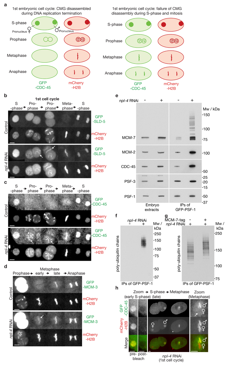
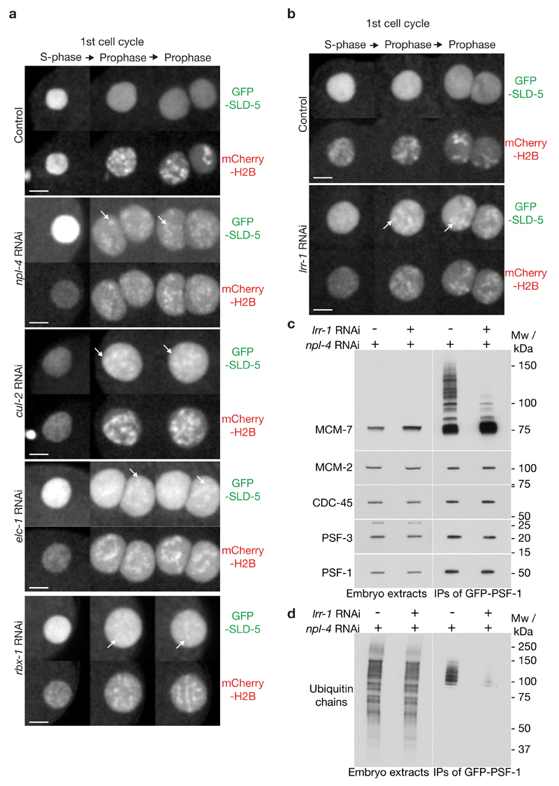
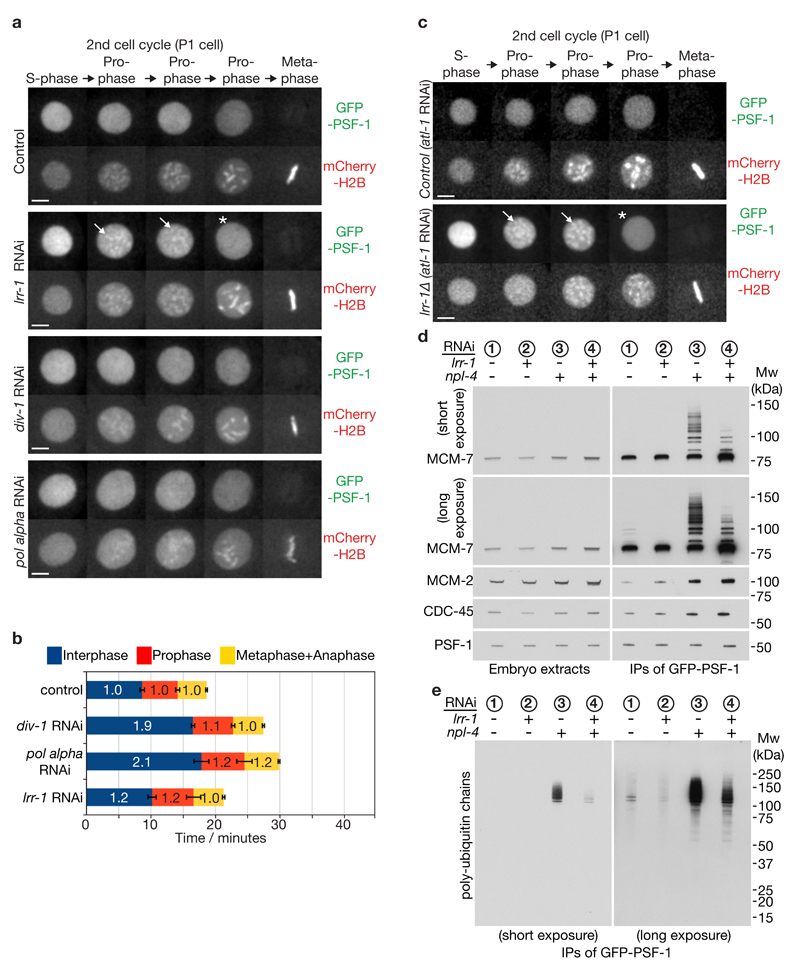
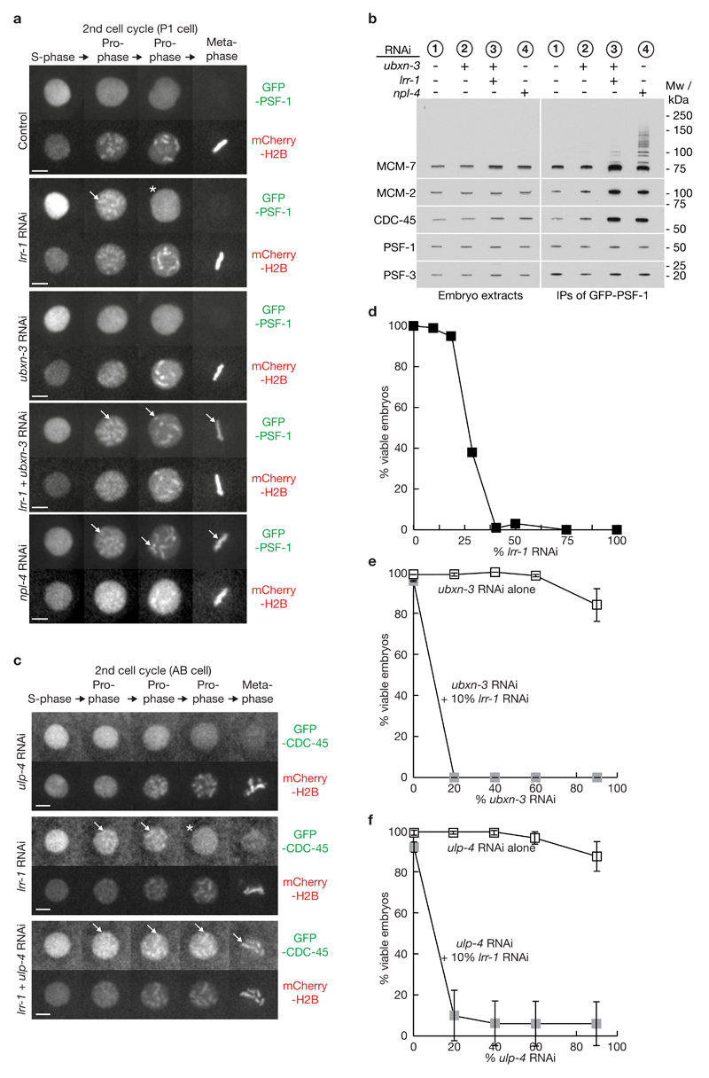
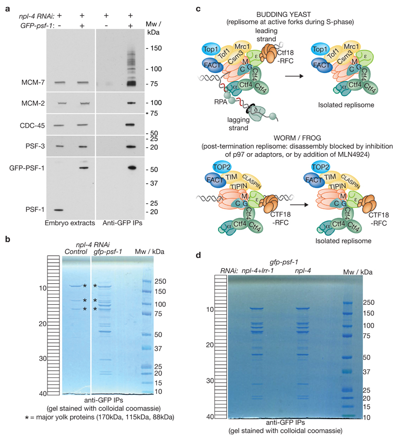
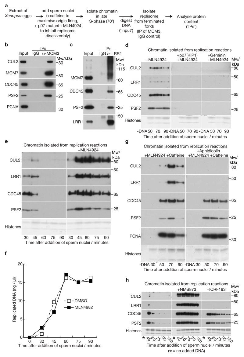
Replisome disassembly is the final step of DNA replication in eukaryotes, involving the ubiquitylation and CDC48-dependent dissolution of the CMG helicase (CDC45-MCM-GINS). Using Caenorhabditis elegans early embryos and Xenopus laevis egg extracts, we show that the E3 ligase CUL-2LRR-1 associates with the replisome and drives ubiquitylation and disassembly of CMG, together with the CDC-48 cofactors UFD-1 and NPL-4. Removal of CMG from chromatin in frog egg extracts requires CUL2 neddylation, and our data identify chromatin recruitment of CUL2LRR1 as a key regulated step during DNA replication termination. Interestingly, however, CMG persists on chromatin until prophase in worms that lack CUL-2LRR-1, but is then removed by a mitotic pathway that requires the CDC-48 cofactor UBXN-3, orthologous to the human tumour suppressor FAF1. Partial inactivation of lrr-1 and ubxn-3 leads to synthetic lethality, suggesting future approaches by which a deeper understanding of CMG disassembly in metazoa could be exploited therapeutically.


Conflict of interest statement

Competing Financial Interests

The authors confirm that they have no competing financial interests.

Figures

Figure 1
Figure 1. The CDC-48 co-factor NPL-4 is required for CMG helicase disassembly during S-phase in the C. elegans early embryo.
(a) Illustration of a live-embryo assay for CMG helicase disassembly, comparing control embryos (‘normal CMG disassembly’) with mutant embryos (‘defective CMG disassembly’). Note that the two nuclei derived from oogenesis and spermatogenesis – referred to in this manuscript as the female and male pronuclei - move together during prophase of the first cell cycle. Following nuclear envelope breakdown, the ‘male’ and ‘female’ sets of chromosomes then intermingle during metaphase. (b) Timelapse video microscopy of the first cell cycle in embryos expressing GFP-SLD-5 and mCherry-HistoneH2B, either untreated or exposed to npl-4 RNAi. The female pronucleus is shown during S-phase, before convergence with the male pronucleus. Prophase begins during migration of the pronuclei. The arrows indicate examples of persistence of GFP-SLD-5 on chromatin during prophase after depletion of NPL-4. (c) Equivalent analysis for embryos expressing GFP-CDC-45. (d) Equivalent data for embryos expressing GFP-MCM-3. The arrow indicates the small pool of GFP-MCM-3 that remains on chromatin during early metaphase after depletion of NPL-4. (e) Homozygous GFP-psf-1 / GFP-psf-1 worms were exposed to npl-4 RNAi or left untreated. Embryos were then isolated and used to generate whole-embryo extracts, before immunoprecipitation of GFP-PSF-1. The indicated proteins were monitored by immunoblotting. (f) The same samples were separated in a 4-12% gradient gel, before immunoblotting with an antibody to poly-ubiquitin chains. (g) Equivalent npl-4 RNAi experiment comparing control worms with homozygous mcm7-5FLAG-9His embryos generated by CRISPR-Cas9. The samples were separated in a 3-8% gradient gel, before immunoblotting with antibody to poly-ubiquitin chains. (h) Timelapse video microscopy of an npl-4 RNAi embryo expressing GFP-CDC-45 and mCherry-HistoneH2B. The GFP signal in the female pronucleus was photo-bleached during early S-phase and then monitored in the subsequent mitosis. Lack of recovery of the GFP signal on ‘female’ chromosomes, compared to the unbleached control male pronucleus, indicated that GFP-CDC45 persists on chromatin after S-phase rather than being reloaded, in embryos lacking NPL-4. The scale bars correspond to 5µm. Unprocessed scans of key immunoblots are shown in Supplementary Figure 8.
Figure 2
Figure 2. CUL-2LRR-1 is required for CMG helicase disassembly during S-phase in C. elegans.
(a-b) Embryos from GFP-sld-5 mCherry-H2B worms were exposed to the indicated RNAi and processed as in Figure 1b. Timelapse images are shown from S-phase to mid-prophase. Five embryos were examined for each treatment and all behaved equivalently. Arrows denote examples of persistence of GFP-SLD-5 on prophase chromatin and scale bars correspond to 5µm. (c-d) Embryos from homozygous GFP-psf-1 / GFP-psf-1 worms were exposed to the indicated RNAi and processed as in Figure 1e-f. Unprocessed scans of key immunoblots are shown in Supplementary Figure 8.
Figure 3
Figure 3. A mitotic pathway for CMG helicase disassembly is revealed in the absence of CUL-2LRR-1.
(a) Embryos from GFP-psf-1 mCherry-H2B worms were exposed to the indicated RNAi treatments, or empty vector in the control, and then processed as in Figure 1b, except that the figure depicts data from the second embryonic cell cycle (P1 cell). Timelapse images are shown from S-phase to metaphase. GFP-PSF1 initially persists on prophase chromatin following depletion of LRR-1 (the arrows denote examples), before being released in late prophase (indicated by asterisk). Scale bars correspond to 5µm. (b) The duration of the indicated cell cycle phases for the experiment in (a) were measured as described in Methods. The data are expressed relative to the length of the corresponding period in control embryos, and represent the mean values (n = 5 embryos; the lines on the boundary of each cell cycle phase indicate standard deviations from the mean). (c) Worms homozygous for GFP-psf-1 and lrr-1Δ were grown in parallel to the equivalent heterozygote (control), as described in Methods. After exposure to atl-1 RNAi (this allows homozygous lrr-1Δ germ cells to proceed with meiosis), the resultant embryos were processed as above. The images depict the second embryonic cell cycle (P1 cell), showing persistent association of GFP-PSF-1 with chromatin during prophase (arrows), before release in late prophase (asterisk). (d-e) Homozygous GFP-psf-1 worms were exposed to the indicated RNAi. Embryos were then isolated and processed as in Figure 1e-f. Unprocessed scans of key immunoblots are shown in Supplementary Figure 8.
Figure 4
Figure 4. The mitotic CMG helicase disassembly pathway requires UBXN-3 and is modulated by the SUMO protease ULP-4, both of which become essential when LRR-1 is depleted.
(a) Embryos from GFP-psf-1 mCherry-H2B worms were exposed to the indicated RNAi and processed as in Figure 3a. The arrows indicate persistent association of GFP-PSF1 with mitotic chromatin (throughout mitosis in the case of RNAi to npl-4, or after simultaneous RNAi to lrr-1 + ubxn-3), whereas the asterisk denotes release of GFP-PSF-1 from chromatin in late prophase in embryos treated only with lrr-1 RNAi. The scale bars correspond to 5µm. (b) Homozygous GFP-psf-1 worms were exposed to the indicated RNAi and isolated embryos were then processed as in Figure 1e. (c) Embryos from GFP-cdc-45 mCherry-H2B worms were exposed to the indicated RNAi and processed as above. The data correspond to the AB cell in the second cell cycle, in which lrr-1 ulp-4 double RNAi leads to persistence of GFP-CDC-45 until at or after nuclear envelope breakdown (8 of 9 embryos tested). (d) Worms were fed on plates where the indicated proportion of bacteria expressed lrr-1 double-stranded RNAi, and embryonic viability was measured as described in Methods (for each timepoint, 69-94 embryos were examined from five adult worms). (e) Worms were fed on the indicated proportion of bacteria expressing ubxn-3 RNAi, either alone or in combination with 10% bacteria expressing lrr-1 RNAi. The data represent the mean values (n = 3 independent experiments; for each timepoint, 70-100 embryos were examined from five adult worms), with the indicated standard deviations from the mean value. (f) Similar experiment involving increasing doses of ulp-4 RNAi, with or without 10% lrr-1 RNAi (n = 3 independent experiments; for each timepoint, 70-100 embryos were examined from five adult worms). Unprocessed scans of key immunoblots are shown in Supplementary Figure 8.
Figure 5
Figure 5. Isolation of the post-termination worm replisome.
(a) Control or homozygous GFP-psf-1 worms were exposed to npl-4 RNAi before being processed as described above for Figure 4. The purified samples were monitored by SDS-PAGE and immunoblotting of the indicated components of the CMG helicase. (b) The remainder of the samples were then resolved in a 4-12% gradient gel, which was stained with colloidal coomassie. The major contaminants in both samples (marked with asterisks) represent the four major yolk proteins of the worm early embryo. Each lane was cut into 40 bands as indicated, before analysis of protein content by mass spectrometry (see Supplementary Table 2). (c) Comparison of the replisome isolated from active replication forks in budding yeast , , , with the isolated post-termination replisome from worm and frog (this study). For simplicity, some of the proteins that act at forks, but that are not present in the isolated replisome, have been omitted. In addition, Mcm10 has been excluded, since its status at forks and its association with the isolated replisome remain unclear (absent from isolated yeast and worm replisomes under physiological conditions, but co-purifying with frog MCM-3 from digested chromatin post-termination). (d) Comparison of isolated replisome material for the experiment in Supplementary Table 3 (worms treated with npl-4 RNAi or npl-4 lrr-1 double RNAi). Unprocessed scans of key immunoblots are shown in Supplementary Figure 8.
Figure 6
Figure 6. CUL2LRR1 associates with the post-termination vertebrate replisome and is recruited to chromatin during DNA replication termination in Xenopus egg extracts.
(a) Experimental scheme for isolation of proteins associated with the CMG helicase after termination in the absence of replisome disassembly, in extracts of Xenopus laevis eggs. (b) Immunoblots of input and the indicated IP samples for the experiment in (a). (c) Replisome disassembly was inhibited with the p97 inhibitor NMS873, and LRR1 was then isolated from digested chromatin at the 70’ timepoint, in parallel with a control IP with IgG, before detection of the indicated proteins by immunoblotting. (d) Chromatin association of the indicated factors was monitored by immunoblotting, at the indicated timepoints after addition of sperm chromatin to egg extracts (except for the -DNA sample that lacked chromatin). Where indicated, replication initiation was blocked by addition of p27(KIP1) or Geminin. The neddylase inhibitor MLN4924 was added to all samples to block replisome disassembly. (e) Timecourse experiment comparing chromatin-bound factors in the absence or presence of the neddylation inhibitor MLN4924. (f) Replication kinetics were monitored for the experiment in (e), by incorporation of radiolabelled α-dATP into newly synthesised DNA (see also Supplementary Figure 5b; source data for repeats of this experiment are included in Supplementary Table 6). (g) Inhibition of DNA synthesis blocks association of CUL2LRR1 with chromatin. DNA synthesis was inhibited with the DNA polymerase inhibitor aphidicolin. Caffeine was added to inactivate the S-phase checkpoint, which otherwise would have reduced the level of CMG on chromatin +Aphidicolin. (h) Analogous experiment to that in (e), showing that CUL2-LRR1 accumulated on chromatin with CMG when replisome disassembly was blocked by the p97 inhibitor NMS873, but chromatin recruitment of CUL2-LRR1 was inhibited if DNA replication termination was delayed by addition of the TOPO2 inhibitor ICRF193. Unprocessed scans of key immunoblots are shown in Supplementary Figure 8.
Figure 7
Figure 7. Active CUL2LRR1 is required for extraction of CMG components from chromatin during DNA replication termination in Xenopus egg extracts.
(a) Experimental scheme. (b) Replication reactions were performed in the presence of MLN4924 to stabilise CUL2LRR1 on chromatin during DNA replication termination in mock-depleted extracts (treated with two rounds of IgG-beads). In contrast, neither CUL2 nor LRR1 were detected on chromatin in CUL2-depleted extracts, confirming the efficiency of the depletion. (c) Depletion of CUL2 also removes LRR1 from the extract (the panel shows immunoblots of the antibody-coupled beads after each of the two rounds of depletion). (d) Kinetics of DNA synthesis in extracts subjected to two rounds of immunoprecipitation with control IgG (‘mock depletion’) or with antibodies to Hs_CUL2-RBX1 (‘CUL2 depletion’, see Methods). Source data for repeats of this experiment are included in Supplementary Table 6. (e) In an analogous experiment, replication reactions were performed in ‘mock-depleted’ and CUL-depleted extracts. A pulse of α-dATP was added for 3’ at either the 60’ or 120’ timepoints, and the incorporation of radiolabel into nascent DNA was monitored after isolation of total DNA, indicating that replication proceeded and completed with similar kinetics in both extracts, consistent with the data in (d). (f) Kinetics of chromatin association of the indicated factors for the same experiment shown in (a-b). Note that the MCM7 immunoblot is over-exposed in order to reveal the ubiquitylated forms of the protein. (g) Mock-depleted or CUL2-depleted extracts were supplemented with the indicated recombinant proteins (X.l. LRR1, wt/mutant Hs_CUL2-RBX1 – see Methods), and chromatin was isolated from the 120’ timepoint in a similar experiment to that described above. Unprocessed scans of key immunoblots are shown in Supplementary Figure 8.

Comment in

  • Two paths to let the replisome go.
    D'Angiolella V, Guardavaccaro D. D'Angiolella V, et al. Cell Death Differ. 2017 Jul;24(7):1140-1141. doi: 10.1038/cdd.2017.75. Epub 2017 May 19. Cell Death Differ. 2017. PMID: 28524853 Free PMC article. No abstract available.

References

    1. Gambus A, et al. GINS maintains association of Cdc45 with MCM in replisome progression complexes at eukaryotic DNA replication forks. Nat Cell Biol. 2006;8:358–366. - PubMed
    1. Moyer SE, Lewis PW, Botchan MR. Isolation of the Cdc45/Mcm2-7/GINS (CMG) complex, a candidate for the eukaryotic DNA replication fork helicase. Proc Natl Acad Sci U S A. 2006 - PMC - PubMed
    1. Bell SP, Labib K. Chromosome Duplication in Saccharomyces cerevisiae. Genetics. 2016;203:1027–1067. - PMC - PubMed
    1. Deegan TD, Diffley JF. MCM: one ring to rule them all. Curr Opin Struct Biol. 2016;37:145–151. - PubMed
    1. O'Donnell M, Li H. The Eukaryotic Replisome Goes Under the Microscope. Curr Biol. 2016;26:R247–256. - PMC - PubMed

Publication types

MeSH terms