

Review
doi: 10.1038/oncsis.2017.14.
Wnt signaling in triple-negative breast cancer
Affiliations
- PMID: 28368389
- PMCID: PMC5520491
- DOI: 10.1038/oncsis.2017.14
Item in Clipboard
Review
Wnt signaling in triple-negative breast cancer
Oncogenesis.
.
Abstract
Wnt signaling regulates a variety of cellular processes, including cell fate, differentiation, proliferation and stem cell pluripotency. Aberrant Wnt signaling is a hallmark of many cancers. An aggressive subtype of breast cancer, known as triple-negative breast cancer (TNBC), demonstrates dysregulation in canonical and non-canonical Wnt signaling. In this review, we summarize regulators of canonical and non-canonical Wnt signaling, as well as Wnt signaling dysfunction that mediates the progression of TNBC. We review the complex molecular nature of TNBC and the emerging therapies that are currently under investigation for the treatment of this disease.
Conflict of interest statement
The authors declare no conflict of interest.
Figures
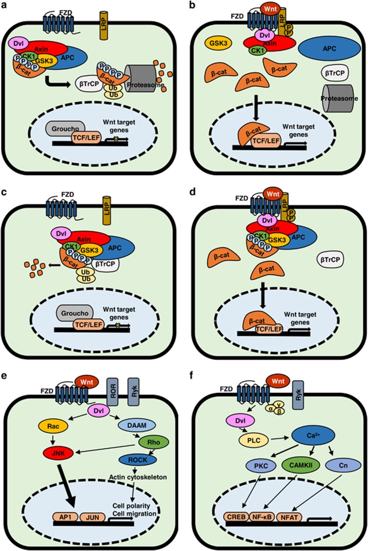
Classical and new Wnt/β-catenin pathway canonical and non-canonical pathways. (a) Overview of the ‘classical’ model of Wnt/β-catenin signaling in OFF state with no ligand bound to FZD receptor. (b) Overview of the ‘classical’ model of Wnt/β-catenin signaling pathway in ON state where Wnt ligand is bound to FZD receptor. (c) Overview of ‘new’ model of Wnt/β-catenin signaling in OFF state with no ligand bound to FZD receptor. (d) Overview of the ‘new’ model of Wnt/β-catenin signaling in ON state with Wnt ligand bound to FZD receptor. (e) Overview of Wnt planar cell polarity (PCP) pathway in ON state. Wnt binds multiple receptors including FZD and co-receptors ROR and Ryk. This activates Rho and Rac, which activate ROCK and c-Jun N-terminal kinase (JNK), respectively, leading to actin polymerization and regulates cytoskeletal arrangements. (f) Overview of Wnt/Ca2+ pathway in ON state. Wnt is able to bind FZD, Ryk to initiate signal transduction, which is effected through Dvl and G proteins (α, β, γ). Gene transcription is induced through proteins PKC, CaMKII and Cn (Calcineurin)-activating transcription factors.
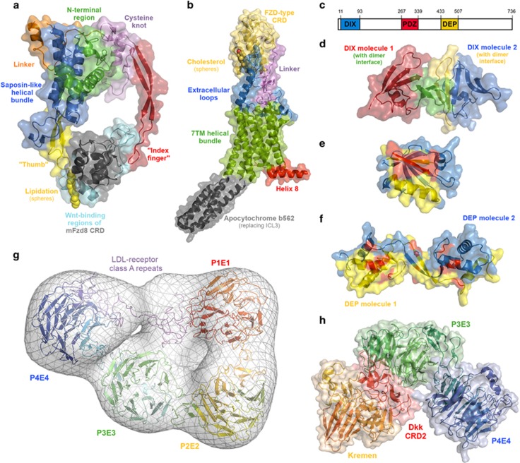
Molecular structures of the key Wnt signaling proteins and interactions. (a) X-ray crystal structure of the Xenopus Wnt8 complex with the mouse FZD8 cysteine-rich domain (PDB 4F0A). The key structural regions of the Wnt fold are highlighted, as are the major Wnt-interacting regions of the CRD. (b) X-ray crystal structure of the Smoothened receptor (PDB 5L7D), a Class F G protein-coupled receptor, related to FZD. The key structural regions of Smo are highlighted, as well as helix 8, which is of relevance for Dishevelled binding by FZD. (c) Schematic representation of the location of the DIX, PDZ and DEP domains within Dvl. (d) X-ray crystal structure of the DIX homodimer (PDB 4WIP). (e) X-ray crystal structure of the PDZ domain bound to a peptide (red; PDB 3CBX). The peptide-binding site is shown in yellow. (f) X-ray crystal structure of a DEP homodimer (PDB 5LNP), highlighting residues known to affect Wnt signaling (shown in red). (g) Model of the LRP6 ectodomain generated by molecular dynamics flexible fitting of the crystal structures of the P1E1–P2E2 domains (PDB 3S94) and P3E3–P4E4 domains (PDB 4A0P), and a homology model of the LDL-R type A domains (generated in Prime, based on the crystal structure of the LDL receptor ectodomain (PDB 1N7D)) to the electron microscopy structure (EMDatabank accession 1964). Gaps in the crystal structures and between the various components modeled using Prime. (h) X-ray crystal structure complex of the cysteine-rich domain 2 of Dickkopf with Kremen and the LRP6 P3E4–P4E4 domains (PDB 5FWW).
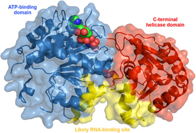
X-ray crystal structure of the ATP-binding and C-terminal helicase domains of the DEAD-box helicase DDX3 (PDB 5E7M). AMP-PNP, a non-hydrolyzable ATP analog, is shown in spheres in the ATP-binding site.
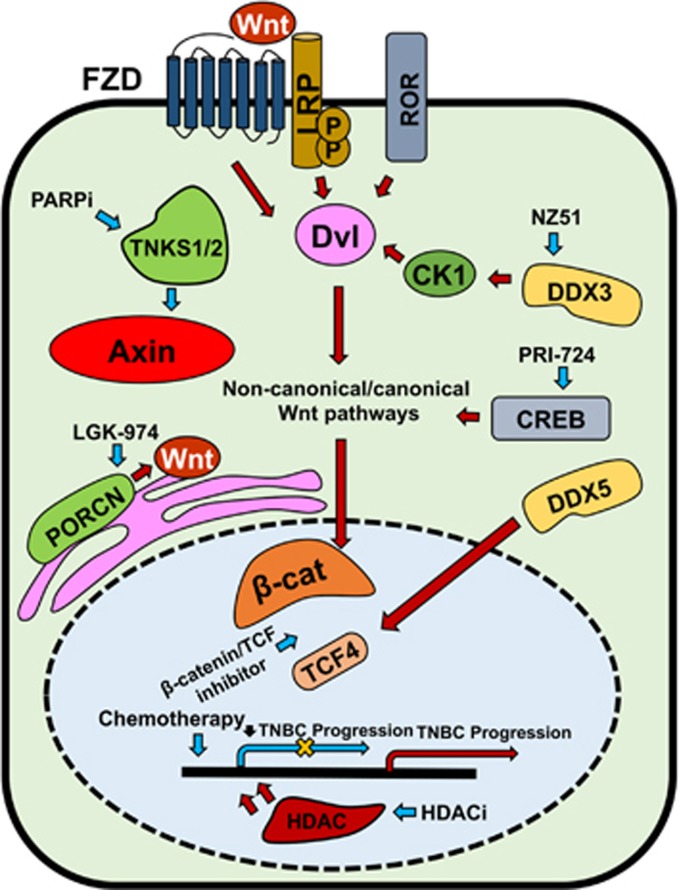
Overview of Wnt signaling regulators contributing to TNBC progression and their targeted therapies. Canonical and non-canonical Wnt pathways are activated through Fzd, LRP and ROR receptors. Blue arrows indicate suppression/inhibition of Wnt regulators and pathways (with a net result of downregulation of Wnt target gene transcription, indicated by yellow cross); red arrows indicate activation of Wnt regulators and pathways.
References
Publication types
LinkOut - more resources
Full Text Sources
Other Literature Sources
