. 2017 May 1;215(9):1396-1406.
doi: 10.1093/infdis/jix138.

Enhanced Glycolytic Metabolism Contributes to Cardiac Dysfunction in Polymicrobial Sepsis

Affiliations

Enhanced Glycolytic Metabolism Contributes to Cardiac Dysfunction in Polymicrobial Sepsis

Zhibo Zheng et al. J Infect Dis. .

Abstract

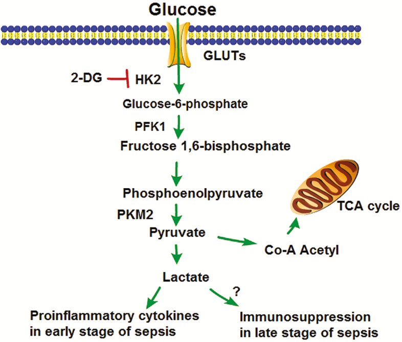
Background: Cardiac dysfunction is present in >40% of sepsis patients and is associated with mortality rates of up to 70%. Recent evidence suggests that glycolytic metabolism plays a critical role in host defense and inflammation. Activation of Toll-like receptors on immune cells can enhance glycolytic metabolism. This study investigated whether modulation of glycolysis by inhibition of hexokinase will be beneficial to septic cardiomyopathy.

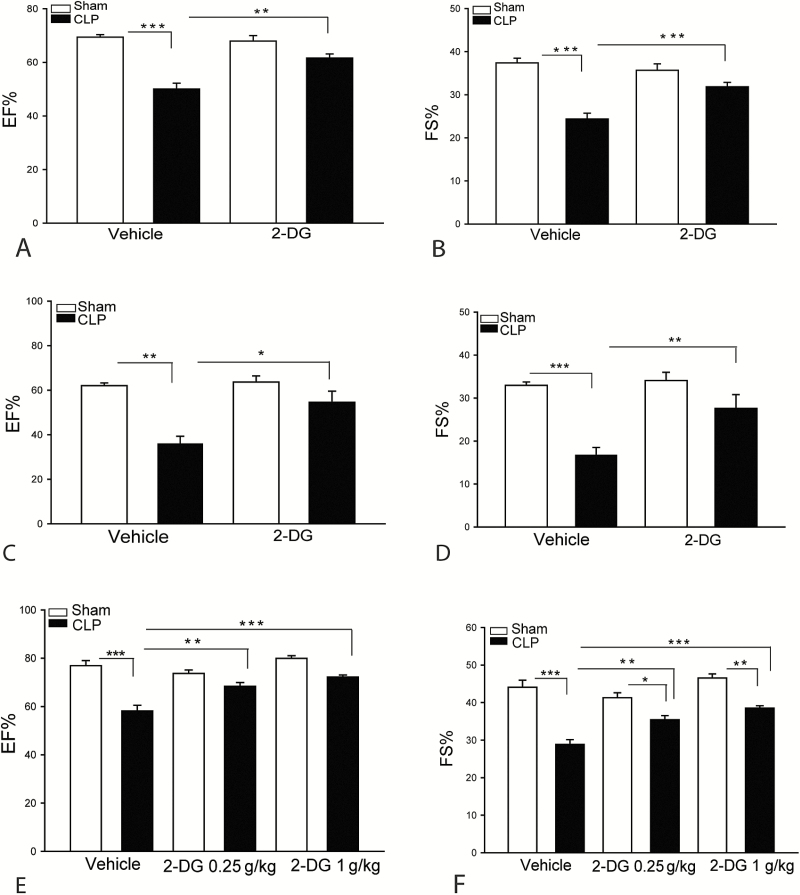
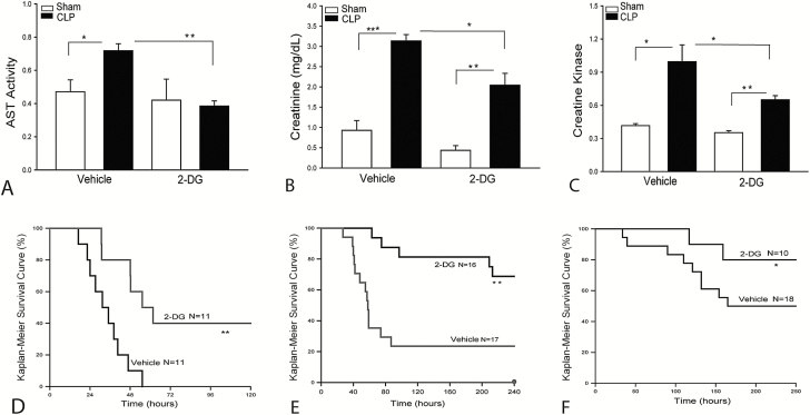
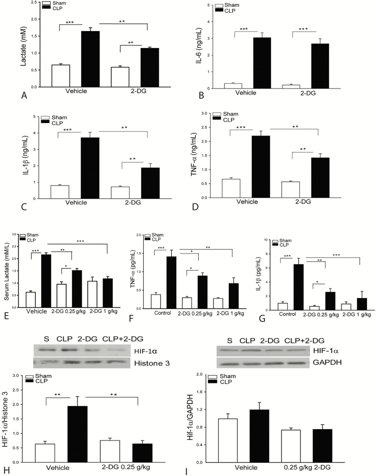
Methods: Male C57B6/J mice were treated with a hexokinase inhibitor (2-deoxy-d-glucose [2-DG], 0.25-2 g/kg, n = 6-8) before cecal ligation and puncture (CLP) induced sepsis. Untreated septic mice served as control. Sham surgically operated mice treated with or without the 2-DG inhibitor served as sham controls. Cardiac function was assessed 6 hours after CLP sepsis by echocardiography. Serum was harvested for measurement of inflammatory cytokines and lactate.

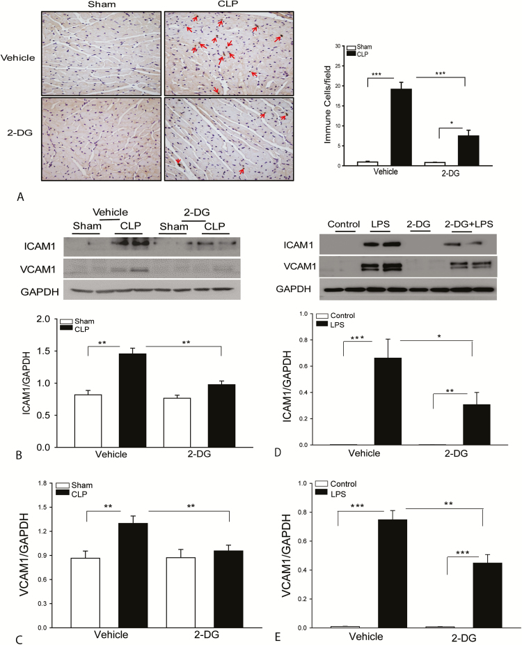
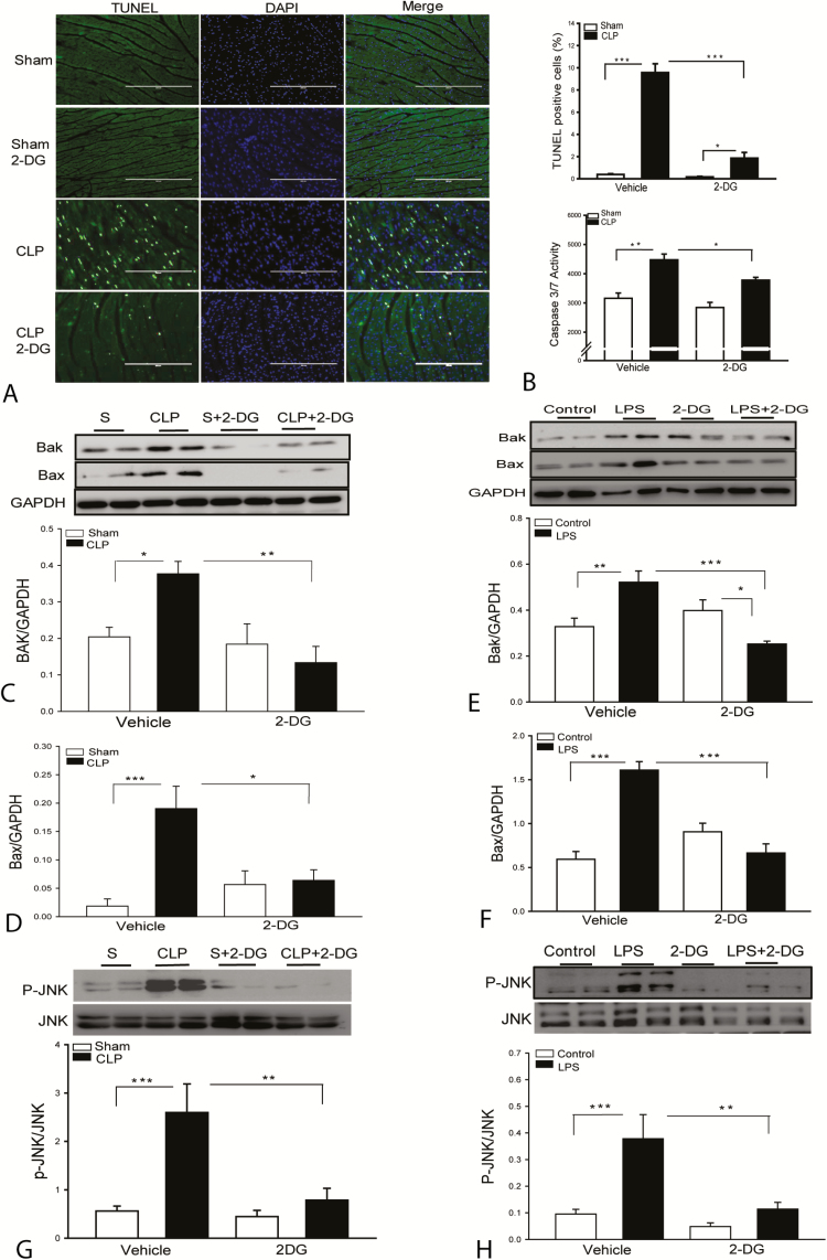
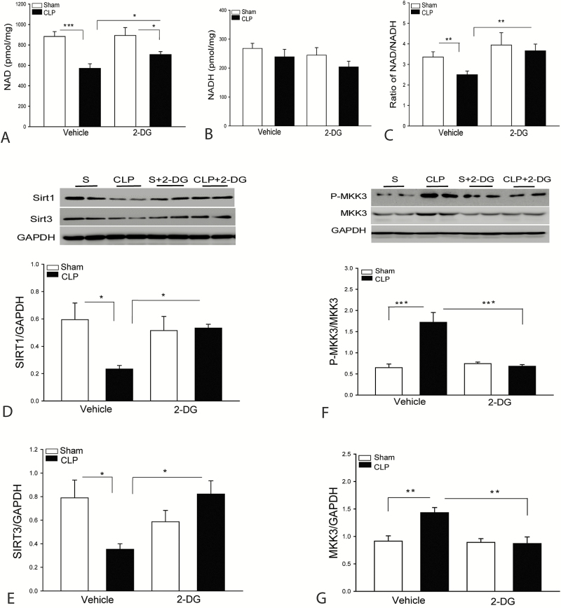
Results: Sepsis-induced cardiac dysfunction was significantly attenuated by administration of 2-DG. Ejection fraction and fractional shortening in 2-DG-treated septic mice were significantly (P < .05) greater than in untreated CLP mice. 2-DG administration also significantly improved survival outcome, reduced kidney and liver injury, attenuated sepsis-increased serum levels of tumor necrosis factor α and interleukin 1β as well as lactate, and enhanced the expression of Sirt1 and Sirt3 in the myocardium, which play an important role in mitochondrial function and metabolism. In addition, 2-DG administration suppresses sepsis-increased expression of apoptotic inducers Bak and Bax as well as JNK phosphorylation in the myocardium.

Conclusions: Glycolytic metabolism plays an important role in mediating sepsis-induced septic cardiomyopathy. The mechanisms may involve regulation of inflammatory response and apoptotic signaling.

Keywords: 2-deoxy-D-glucose; cardiomyopathy; glycolysis; inflammatory responses.; sepsis.


Figures

Figure 1.
Figure 1.
Inhibition of hexokinase attenuates sepsis-induced cardiac dysfunction. Mice were treated with a single dose of 2-deoxy-d-glucose (2-DG) (2 g/kg) 3 hours prior to cecal ligation and puncture (CLP). Cardiac function was examined by echocardiography 6 hours after CLP. A, Ejection fraction (EF%). B, Fractional shortening (FS%). C and D, 2-DG administration at 0.5 g/kg body weight also significantly attenuated CLP sepsis-induced cardiac dysfunction. E and F, 2-DG treatment at 0.25 and 1 g/kg also significantly improves cardiac function. n = 6–8/group. *P < .05, **P < .01, and ***P < .001 compared with indicated groups.
Figure 2.
Figure 2.
Administration of 2-deoxy-d-glucose (2-DG) attenuates sepsis-induced organ injury and improves survival outcome. Mice were treated with 2-DG (0.25 g/kg body weight) 3 hours before induction of cecal ligation and puncture (CLP) sepsis. Serum levels of aspartate aminotransferase (AST; A), creatinine (B), and creatine kinase (C) were measured by commercially available enzyme-linked immunosorbent assay kits. D–F, 2-DG administration improves survival outcome. Mice were treated with 2-DG (2 g/kg body weight [D] or 0.25 g/kg body weight [E]) 3 hours prior to CLP and were then re-treated with 2-DG (500 mg/kg) 24 hours after CLP. D and E, Acute CLP sepsis. F, Chronic CLP septic mice treated with 2-DG at 0.5 kg/kg body weight 3 hours prior to CLP. The animals were monitored for lethality every 2 hours for up to 250 hours following CLP. n = 10–18/group. *P < .05, **P < .01, and ***P < .001 compared with indicated groups.
Figure 3.
Figure 3.
Administration of 2-deoxy-d-glucose (2-DG) attenuates sepsis-induced accumulation of immune cells and prevents intercellular cell adhesion molecule 1 (ICAM-1) and vascular cell adhesion molecule 1 (VCAM-1) expression in the myocardium. A, Eight hours after cecal ligation and puncture (CLP)–induced sepsis, hearts were harvested and sectioned for immunostaining of immune cells with specific antibody NIMP-R1411b, which can recognize Ly-6G and Ly-6C neutrophils, monocytes, and macrophages. 2-DG prevents sepsis-induced increases in the expression of ICAM-1 (B) and VCAM-1 (C). n = 4–6 per group. 2-DG treatment attenuated lipopolysaccharide (LPS)–induced ICAM-1 (D) and VCAM-1 (E) expression in endothelial cells. Human umbilical vein endothelial cells were treated with LPS (1 µg/mL) in the presence or absence of 2-DG. The cells were harvested 12 hours after treatment. Cellular proteins were prepared for immunoblotting. n = 8 replicates in each group. *P < .05, **P < .01, and ***P < .001 compared with indicated groups.
Figure 4.
Figure 4.
Administration of 2-deoxy-d-glucose (2-DG) attenuates sepsis-induced lactate and inflammatory cytokine production. Serum was harvested 8 hours after cecal ligation and puncture (CLP) sepsis. The levels of lactate (A), tumor necrosis factor (TNF-α; B), interleukin 1β (IL-1β; C), and interleukin 6 (IL-6; D) were analyzed with enzyme-linked immunosorbent assay. Administration of 2-DG at 0.25 or 1 g/kg body weight also significantly reduced serum lactate (E), TNF-α (F), and IL-1β (G). 2-DG (0.25 g/kg) attenuated sepsis-induced increases in the nuclear hypoxia-inducible factor (HIF-1α) levels (H). CLP sepsis did not significantly alter cytosolic HIF-1α levels in the myocardium (I). n = 4–6/group. *P < .05, **P < .01, and ***P < .001 compared with indicated groups.
Figure 5.
Figure 5.
Administration of 2-deoxy-d-glucose (2-DG) attenuates sepsis-induced myocardial apoptosis. A, Hearts were harvested 8 hours after cecal ligation and puncture (CLP) sepsis and sectioned for TUNEL assay. B, Myocardial caspase-3/7 activity was analyzed by enzyme-linked immunosorbent assay. 2-DG treatment prevents sepsis-induced Bak (C) and Bax (D) expression in the myocardium. 2-DG treatment prevents lipopolysaccharide (LPS)–induced Bak (E) and Bax (F) expression in endothelial cells. n = 8 replicates/group. 2-DG treatment prevents sepsis-induced JNK phosphorylation in the myocardium (G; n = 4–6/group) and LPS-increased JNK phosphorylation in the endothelial cells (H). Human umbilical vein endothelial cells were treated with lipopolysaccharide (1 µg/mL) in the presence or absence of 2-DG. The cells were harvested 12 hours after treatment. Cellular proteins were prepared for immunoblotting. n = 8 replicates/group. *P < .05, **P < .01, and ***P < .001 compared with indicated groups.
Figure 6.
Figure 6.
Administration of 2-deoxy-d-glucose (2-DG) attenuated sepsis-induced decreases in the levels of nicotinamide adenine dinucleotide (NAD+, oxidized form), Sirt1, and Sirt3 in the myocardium. Hearts were harvested 8 hours after cecal ligation and puncture (CLP) for preparation of cellular proteins. NAD+ (A) and nicotinamide adenine dinucleotide (NADH, reduced form) (B) levels were examined by commercially available kits. C, Ratio of NAD+/NADH. D and E, 2-DG treatment increased the levels of Sirt1 (D) and Sirt3 (E) in the myocardium. F and G, 2-DG treatment prevents MKK3 phosphorylation and expression in the myocardium. n = 4–6/group. *P < .05, **P < .01, and ***P < .001 compared with indicated groups.
Figure 7.
Figure 7.
Two-deoxy-d-glucose (2-DG) suppresses HK2 activity, thus reducing lactate production. Abbreviations: GLUT, glucose transport protein; HK2, hexokinase 2; PFK1, phosphofrutokinase 1; PKM2, pyruvate kinase isozymes M1/2; TCA, tricarboxylic acid.

References

    1. Mayr FB, Yende S, Angus DC. Epidemiology of severe sepsis. Virulence 2014; 5:4–11. - PMC - PubMed
    1. Fernandes CJ, Jr, de Assuncao MS. Myocardial dysfunction in sepsis: a large, unsolved puzzle. Crit Care Res Pract 2012; 2012:896430. - PMC - PubMed
    1. Gao M, Ha T, Zhang X, et al. Toll-like receptor 3 plays a central role in cardiac dysfunction during polymicrobial sepsis. Crit Care Med 2012; 40:2390–9. - PMC - PubMed
    1. Feng Y, Zou L, Zhang M, Li Y, Chen C, Chao W. MyD88 and Trif signaling play distinct roles in cardiac dysfunction and mortality during endotoxin shock and polymicrobial sepsis. Anesthesiology 2011; 115:555–67. - PMC - PubMed
    1. Zou L, Feng Y, Chen YJ, et al. Toll-like receptor 2 plays a critical role in cardiac dysfunction during polymicrobial sepsis. Crit Care Med 2010; 38:1335–42. - PMC - PubMed

Publication types

MeSH terms