. 2017 Jun 1;158(6):1929-1938.
doi: 10.1210/en.2017-00061.

Combined Deletion of the Vitamin D Receptor and Calcium-Sensing Receptor Delays Wound Re-epithelialization

Affiliations

Combined Deletion of the Vitamin D Receptor and Calcium-Sensing Receptor Delays Wound Re-epithelialization

Yuko Oda et al. Endocrinology. .

Abstract

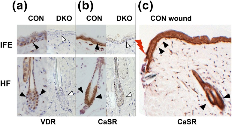
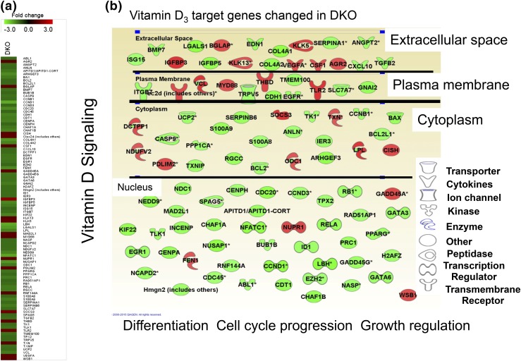
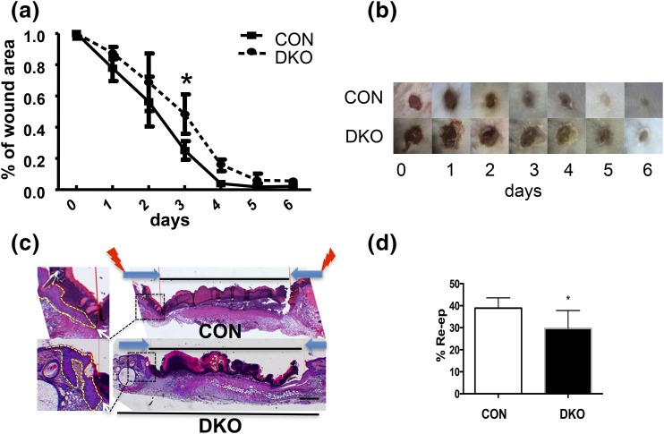
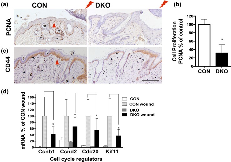
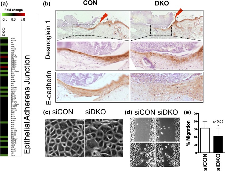
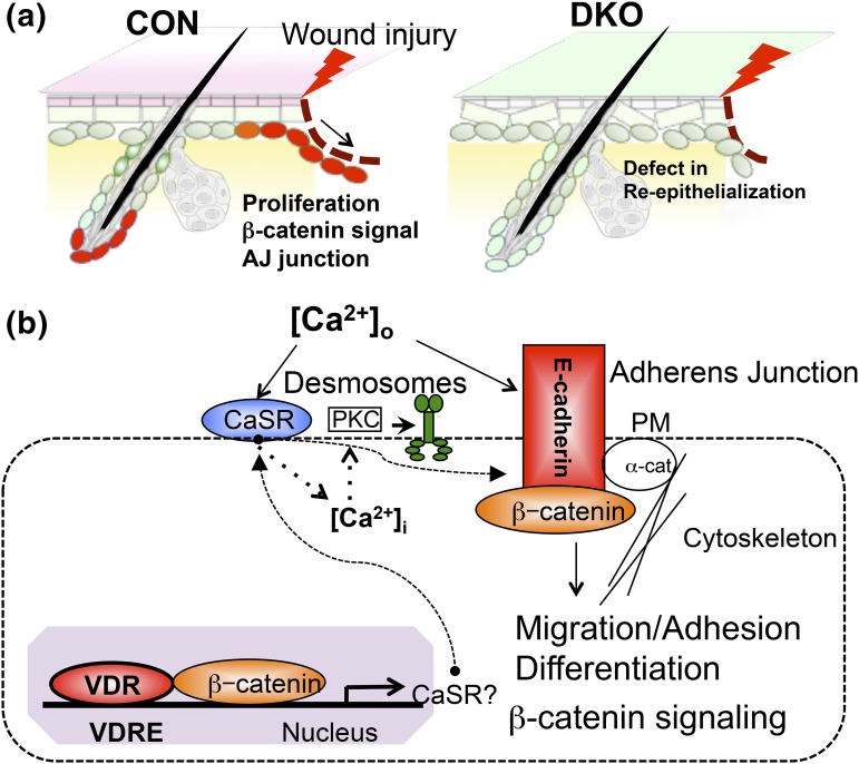
When the skin is injured, keratinocytes proliferate, migrate, and differentiate to regenerate the epidermis. We recently showed that ablation of the vitamin D receptor (Vdr) in keratinocytes delays wound re-epithelialization in mice also fed a low-calcium diet, implicating a cooperative role of Vdr and calcium signaling in this process. In this study, we examined the role of vitamin D and calcium signaling in wound healing by deleting their receptors, Vdr and the calcium-sensing receptor (Casr). Gene expression profiling of neonatal epidermis lacking both Vdr and Casr [Vdr and Casr double knockout (DKO)] specifically in keratinocytes revealed that DKO affects a number of pathways relevant to wound healing, including Vdr, β-catenin, and adherens junction (AJ) signaling. In adult skin, DKO caused a significant delay in wound closure and re-epithelialization, whereas myofibroblast numbers and matrix deposition were unaffected. The injury-induced proliferation of epidermal keratinocytes was blunted in both epidermis and hair follicles, and expression of β-catenin target genes was reduced in the DKO. Expression of E-cadherin and desmoglein 1 was reduced in the shortened leading edges of the epithelial tongues re-epithelializing the wounds, consistent with the decreased migration rate of DKO keratinocytes in vitro. These results demonstrate that Vdr and Casr are required for β-catenin-regulated cell proliferation and AJ formation essential for re-epithelialization after wounding. We conclude that vitamin D and calcium signaling in keratinocytes are required for a normal regenerative response of the skin to wounding.


Figures

Figure 1.
Figure 1.
DKO mice were generated in which both Vdr and Casr were removed from Krt14-expressing keratinocytes. Immunohistochemistry for (a) Vdr and (b, c) Casr in 3-month-old DKO skin (arrowheads) and littermate CON skin. The Casr staining in the epidermis at the edge of the wound (lightning bolt) in CON skin is also shown. Representative images of staining in IFE and HF are shown.
Figure 2.
Figure 2.
Deletion of both Vdr and Casr downregulates Vdr signaling in neonatal epidermis. (a, b) Gene expression profiling of neonatal (days P1 to P3) epidermis lacking both Vdr and Casr (DKO). (a) The heat maps for DKO show the fold changes (KO/CON) for representative genes for vitamin D signaling. (b) The changes in vitamin D3 target genes in DKO epidermis are shown with their subcellular localization and function. The lighter elements denote downregulation, and the darker elements denote upregulation.
Figure 3.
Figure 3.
Deletion of both Vdr and Casr delays re-epithelialization after wounding. (a) Six-mm, full-thickness skin biopsy wounds were made on the backs of 3-month-old DKO mice and their CON littermates. The areas of the wounds were measured 0 to 6 days later and normalized to the original wound area (time 0) in DKO and CON mice. The bars enclose the mean ± standard deviation (SD). *P < 0.05 (n = 7 to 8). (b) Representative 0- to 6-day wound photographs of DKO and CON mice are shown. (c) Three-mm, full-thickness skin biopsy wounds were made on the back skin of 3-month-old DKO and CON mice, and the re-epithelialization was evaluated histologically at day 3 by hematoxylin and eosin staining. Images at higher magnification (boxed, right) show the edge (white arrows) of epithelial tongues (dotted lines, left) from which the distance was measured to evaluate the re-epithelialization. (d) Percent re-epithelialization was quantitatively evaluated by analysis of different cross-sections (n = 6; three mice each). Percent re-epithelialization was defined as the distance traveled by both epithelial margins [large dark arrows in (c), right] divided by the distance needed to travel to fully re-epithelialize the wound [lightning bolts in (c), right]. The bars enclose mean ± SD. Statistical significance was evaluated by t test. *, P < 0.05.
Figure 4.
Figure 4.
Combined deletion of Vdr and Casr blunts injury-induced cell proliferation and activation of β-catenin signaling. (a) PCNA staining at the wounding edge (lightning bolt) of DKO and littermate CON mice (3 days after wounding at 3 months old; arrowhead). The representative images for three analyses are shown. (b) Cell proliferation in IFE was quantified by Bioquant, shown in the bar graph with statistical significance. Mean ± SD shown (n = 6). *P < 0.05. (c) Immunohistochemistry for the β-catenin target gene Cd44 (arrowhead) at wound sections similar to those in (a) for DKO and CON. (d) The mRNA levels for β-catenin–regulated Ccnd1 and other cell cycle regulators in control and wounded skin in DKO and CON mice were evaluated by qPCR. Statistical significance compared with wounded CON skin vs wounded DKO skin is shown by an asterisk (*P < 0.05). Mean ± SD shown (n = 3).
Figure 5.
Figure 5.
Deletion of both Vdr and Casr (DKO) impairs adhesion and migration of keratinocytes to re-epithelize the wound. (a) DKO downregulation of AJ signaling in epidermis as shown in microarray analyses. (b) Immunohistochemistry for desmoglein 1 (upper two panels) and E-cadherin (bottom panels) on the wound sections of CON (left) and DKO (right) mice (3 days after wounding at 3 months of age). The upper panels for desmoglein 1 are of low magnification, with the boxed regions shown in the middle panels at higher magnification. The lightning bolts show wound margins. Representative images from two analyses are shown. (c) The phase-contrast morphology of human keratinocytes transfected by siDKO compared with siCON (phase microscopy). (d, e) Migration capability was assessed by the scratch assay. Confluent cultures of transfected cells were treated to block proliferation, and they were scratched by pipette tips. After 16 hours, the migration rate was quantitated by measuring the amount of open area remaining in the scratch region through Bioquant software, and the results expressed as the ratio of closure after 16 hours to the original area of the scratch. Mean ± SD shown (n = 12). *P < 0.05.
Figure 6.
Figure 6.
A proposed model in which Vdr and calcium signaling concurrently control wound re-epithelialization through regulation of AJ formation and β-catenin signaling. (a) Summary showing deficiency in both Vdr and Casr prevents proliferation and migration of keratinocytes to delay wound (lightning bolt) re-epithelialization of epidermis. (b) A proposed action of vitamin D and calcium signaling, in which they cross-talk to regulate AJ formation, β-catenin signaling through AJ signaling, and desmosome function through calcium-regulated PKCα activity. Both Vdr and Casr concurrently control β-catenin signaling and AJ signaling, which govern adhesion, migration, and differentiation of regenerating keratinocytes at the wound edge.

Similar articles

Cited by

References

    1. Sen CK. Wound healing essentials: let there be oxygen. Wound Repair Regen. 2009;17(1):1–18. - PMC - PubMed
    1. Burkievcz CJ, Skare TL, Malafaia O, Nassif PA, Ribas CS, Santos LR. Vitamin D deficiency in patients with chronic venous ulcers. Rev Col Bras Cir. 2012;39(1):60–63. - PubMed
    1. Zubair M, Malik A, Meerza D, Ahmad J. 25-Hydroxyvitamin D [25(OH)D] levels and diabetic foot ulcer: is there any relationship? Diabetes Metab Syndr. 2013;7(3):148–153. - PubMed
    1. Oda Y, Tu CL, Menendez A, Nguyen T, Bikle DD. Vitamin D and calcium regulation of epidermal wound healing. J Steroid Biochem Mol Biol. 2016;164:379–385. - PMC - PubMed
    1. Luderer HF, Nazarian RM, Zhu ED, Demay MB. Ligand-dependent actions of the vitamin D receptor are required for activation of TGF-β signaling during the inflammatory response to cutaneous injury. Endocrinology. 2013;154(1):16–24. - PMC - PubMed

MeSH terms