. 2017 Jun 1;27(6):3346-3359.
doi: 10.1093/cercor/bhx070.

Occipital White Matter Tracts in Human and Macaque

Affiliations

Occipital White Matter Tracts in Human and Macaque

Hiromasa Takemura et al. Cereb Cortex. .

Abstract

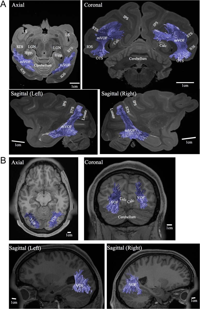
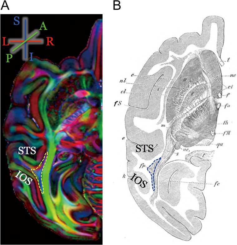
We compare several major white-matter tracts in human and macaque occipital lobe using diffusion magnetic resonance imaging. The comparison suggests similarities but also significant differences in the tracts. There are several apparently homologous tracts in the 2 species, including the vertical occipital fasciculus (VOF), optic radiation, forceps major, and inferior longitudinal fasciculus (ILF). There is one large human tract, the inferior fronto-occipital fasciculus, with no corresponding fasciculus in macaque. We could identify the macaque VOF (mVOF), which has been little studied. Its position is consistent with classical invasive anatomical studies by Wernicke. VOF homology is supported by similarity of the endpoints in V3A and ventral V4 across species. The mVOF fibers intertwine with the dorsal segment of the ILF, but the human VOF appears to be lateral to the ILF. These similarities and differences between the occipital lobe tracts will be useful in establishing which circuitry in the macaque can serve as an accurate model for human visual cortex.

Keywords: comparative study; diffusion MRI; vertical occipital fasciculus; visual cortex; white matter.


Figures

Figure 1.
Figure 1.
Major white-matter tracts with at least one endpoint in occipital cortex. Axial view of major white-matter tracts (ILF, red; IFOF, purple; OR, green; Forceps Major, dark yellow; VOF, blue) overlaid on anatomical image in a representative macaque (A, subject M1, ex vivo) and human (B, subject H1, HCP90 data set). The small panel in the left side in panel A indicates the size of the macaque tracts in an identical spatial scaling with human figure.
Figure 2.
Figure 2.
The position of the mVOF. The position of the mVOF identified in PDD map (subject M1, ex vivo). The color scheme depicts the PDD in each voxel (blue, superior–inferior; green, anterior–posterior; red, left–right). In the axial slice (left panel), we could see the white-matter portion with predominantly superior–inferior diffusion signal between superior temporal sulcus (STS) and inferior occipital sulcus (IOS). In the coronal slice (right panel), this region is located lateral to the calcarine sulcus and the OR (which has anterior–posterior PDD, green). Scale bar (white) in each panel indicates 10 mm. Light blue dotted line in left panel indicates the position of coronal slice in the right panel, vice versa. LS, lateral sulcus; IPS, intraparietal sulcus; Calc, calcarine sulcus; POS, parieto-occipital sulcus; OTS, occipito-temporal sulcus.
Figure 3.
Figure 3.
Human and macaque VOF identified using tractography. (A) mVOF identified using tractography, overlaid on nondiffusion weighted (b = 0) image (subject M1; left top, axial slice; right top, coronal slice; bottom, sagittal slices). Lunate, lunate sulcus; Hipp, hippocampus. Other conventions are identical to those in Figure 2. (B) Human VOF identified using tractography, overlaid on T1-weighted image (subject H1, HCP90 data set). Scale bar (white line) in each panel indicate 10 mm.
Figure 4.
Figure 4.
Statistical evidence supporting the existence of the mVOF. (A) The bootstrapped RMSE distributions for the unlesioned (orange) and lesioned (blue) models. The RMSE is calculated for voxels in the right mVOF in subject M1 (see Material and Methods). The 2 distributions are widely separated (S = 14.03). (B) Two-dimensional histogram comparing the RMSE between unlesioned (abscissa) and lesioned (ordinate) models (right mVOF voxels in M1). Color map indicates the number of voxels. The increased error in the lesioned model arises from a subset of the voxels that have larger RMSE for the lesioned compared with the full model (upper left).
Figure 5.
Figure 5.
Cortical visual maps near VOF endpoints in human and macaque. (A) Cortical maps near the mVOF endpoint (subject M1, right hemisphere). In this plot, we calculated the spatial distance between mVOF endpoints and gray matter voxels, and counted the number of mVOF streamlines having endpoints close to the gray matter voxel (see Materials and Methods). The boundaries between visual areas are manually identified using an MRI-based atlas (Saleem and Logothetis 2012). We observed the putative mVOF termination near V3A and V4d dorsally, as well as V4v and TEO ventrally. See Supplementary Fig. 5 for more examples. (B) Cortical maps near human VOF endpoint in a representative hemisphere from HCP90 data set (subject H1, right hemisphere). The boundaries between cortical areas are estimated using a surface-based probabilistic atlas (Wang et al. 2015). (C) Cortical maps near human VOF endpoints in relation to visual field maps estimated individually using fMRI (Dumoulin and Wandell 2008; STN96 data set, subject H4, right hemisphere). The boundaries between cortical areas are defined by the boundaries of polar angle and eccentricity estimated by fMRI-based visual field mapping (see captions at the center; circular inset; UVM, upper vertical meridian; HM, horizontal meridian; LVM, lower vertical meridian).
Figure 6.
Figure 6.
Human-macaque comparison of VOF position with respect to other tracts. (A) Spatial relationship between the OR (green), the ILF (magenta), and VOF (blue) in human and macaque. Macaque tracts are depicted from 2 different viewpoints (sagittal and coronal). (B) VOF and ILF cortical endpoints in human and macaque in the right hemisphere (human, subject H1 from HCP90 data set; macaque, subject M1), shown as cortical mesh representation from 2 different viewpoints. Color on the mesh indicates the approximate position of the ILF, VOF endpoints, and their overlap (red: ILF, blue: VOF, purple: overlap). See Supplementary Figure 11 for the summary statistics on the estimated overlap.
Figure 7.
Figure 7.
History of the mVOF. The comparison of the mVOF position in modern diffusion MRI data and Wernicke's study (Wernicke 1881). (A) The position of the mVOF in PDD map in the left hemisphere (subject M1). This slice is chosen to match the schematic diagram in Wernicke's study (right, B). While it is impossible to completely match the slice between modern data and classical work, the position of the mVOF (left) and “vertical occipital bundle” (fp, right panel) is qualitatively consistent; both are located between the STS (“e, Parallelfurche” in Wernicke) and IOS (“k, vordere Occipitalfurche” in Wernicke).
Figure 8.
Figure 8.
Schematic diagram of estimated human-macaque occipital fiber system. This diagram schematically describes the position of 3 major white-matter pathways in the right hemisphere in humans (left panel) and macaque (right panel); the OR (green), the ILF (red), and the VOF (blue). As compared with macaques, human VOF is moved to the lateral side of the white matter and becomes relatively distinguishable from the ILF.

References

    1. Aggarwal M, Nauen DW, Troncoso JC, Mori S. 2015. Probing region-specific microstructure of human cortical areas using high angular and spatial resolution diffusion MRI. Neuroimage. 105:198–207. - PMC - PubMed
    1. Ajina S, Pestilli F, Rokem A, Kennard C, Bridge H, Brown EN. 2015. Human blindsight is mediated by an intact geniculo-extrastriate pathway. eLife Sciences. 4:e08935. - PMC - PubMed
    1. Amano K, Wandell BA, Dumoulin SO. 2009. Visual field maps, population receptive field sizes, and visual field coverage in the human MT+ complex. J Neurophysiol. 102:2704–2718. - PMC - PubMed
    1. Arcaro MJ, McMains SA, Singer BD, Kastner S. 2009. Retinotopic organization of human ventral visual cortex. J Neurosci. 29:10638–10652. - PMC - PubMed
    1. Bailey P, Von Bonin G, Davis EW, Garol HW, Mcculloch WS. 1944. Further observations on associational pathways in the brain of macaca mulatta. J Neuropathol Exp Neurol. 3:413.

Publication types