. 2017 Jul 1;27(7):657-668.
doi: 10.1093/glycob/cwx026.

Siglec-8 and Siglec-9 binding specificities and endogenous airway ligand distributions and properties

Affiliations

Siglec-8 and Siglec-9 binding specificities and endogenous airway ligand distributions and properties

Huifeng Yu et al. Glycobiology. .

Abstract

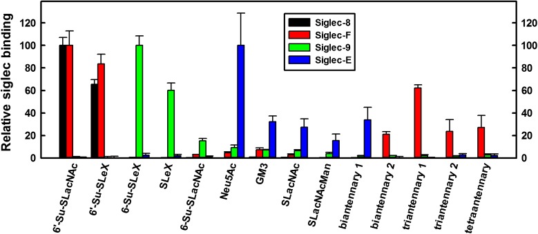
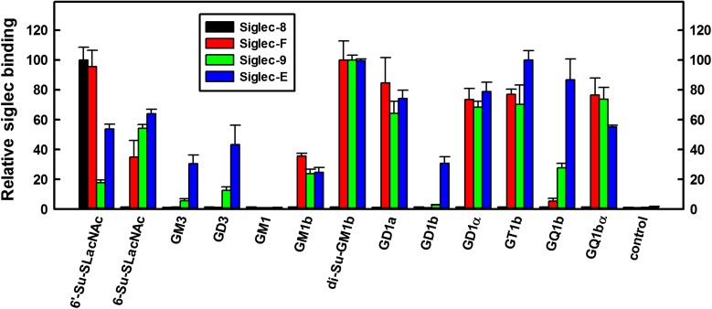
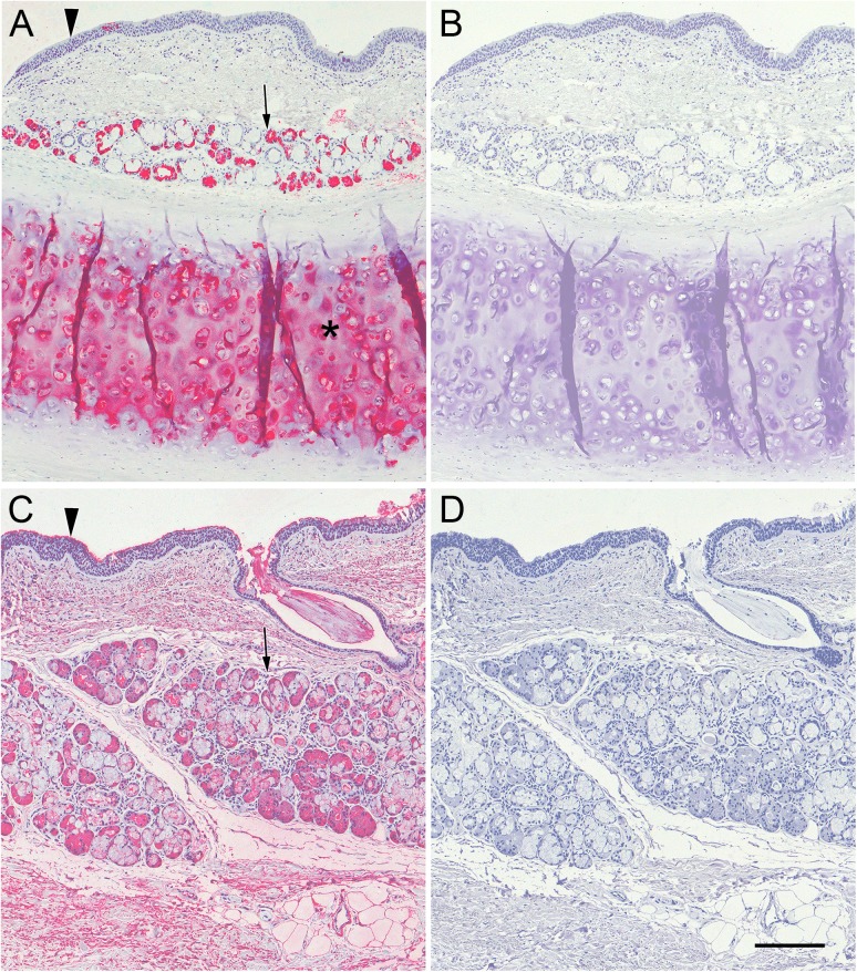
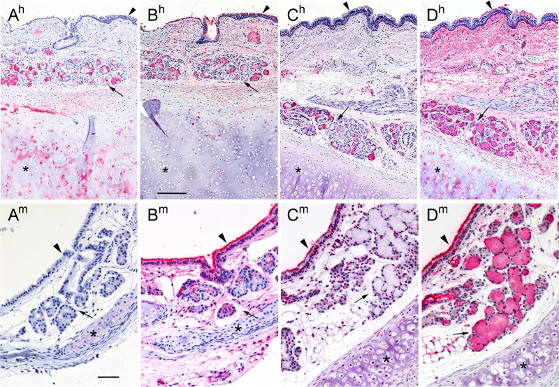
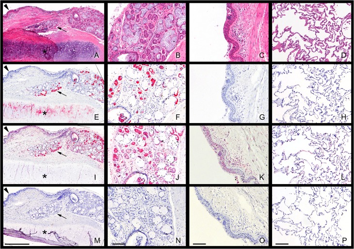
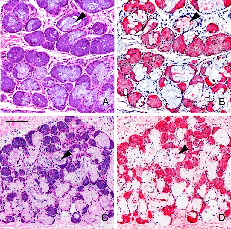
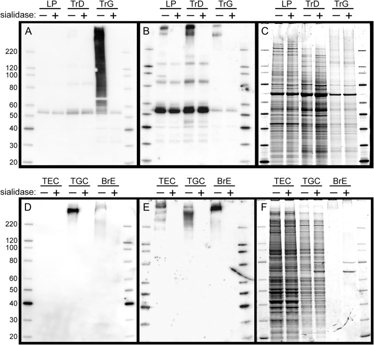
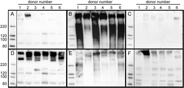
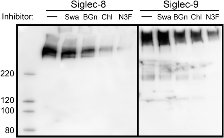
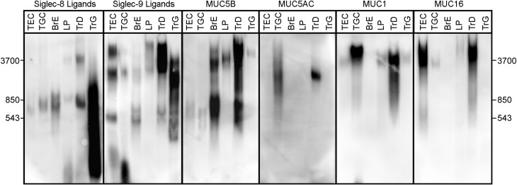
Siglecs are transmembrane sialoglycan binding proteins, most of which are expressed on leukocyte subsets and have inhibitory motifs that translate cell surface ligation into immune suppression. In humans, Siglec-8 on eosinophils, mast cells and basophils and Siglec-9 on neutrophils, monocytes and some T-cells, mediate immune cell death, inhibition of immune mediator release and/or enhancement of anti-inflammatory mediator release. Endogenous sialoglycan ligands in tissues, mostly uncharacterized, engage siglecs on leukocytes to inhibit inflammation. Glycan array analyses demonstrated that Siglec-8, Siglec-9 and their mouse counterparts Siglec-F and Siglec-E (respectively) have distinct glycan binding specificities, with Siglec-8 more structurally restricted. Since siglecs are involved in lung inflammation, we studied Siglec-8 and Siglec-9 ligands in human lungs and airways. Siglec-8 ligands are in tracheal submucosal glands and cartilage but not airway epithelium or connective tissues, whereas Siglec-9 ligands are broadly distributed. Mouse airways do not have Siglec-8 ligands, whereas Siglec-9 ligands are on airways of both species. Extraction of human airways and lung followed by electrophoretic resolution and siglec blotting revealed Siglec-8 ligands in extracts of human trachea and cultured tracheal gland cells, but not parenchyma or cultured airway epithelial cells whereas Siglec-9 ligands were extracted from all airway and lung tissues and cells tested. Siglec-8 and Siglec-9 ligands in airways appear to be high molecular weight O-linked sialoglycoproteins. These data reveal differential glycan specificities of Siglec-8, Siglec-9 and their mouse counterparts Siglec-F and Siglec-E, and the tissue distributions and molecular characteristics of Siglec-8 and Siglec-9 sialoglycan ligands on human airways and lungs.

Keywords: airways; sialic acid; siglec; submucosal glands; trachea.


Figures

Fig. 1.
Fig. 1.
Binding of human Fc chimeras of Siglec-8, -F, -9 and -E to the Consortium for Functional Glycomics (CFG) glycan array. Siglec-human Fc chimeras were overlaid on the CFG printed microarray version 5.1 (610 glycans, http://glycomics.scripps.edu/CoreH/CoreHarray070112V5.1.pdf) and binding detected using fluorescently labeled anti-human IgG secondary antibody. Binding of each siglec is normalized to its maximum binding glycan. The 14 sialoglycans that supported ≥15% of maximum binding to any of the four siglecs are included in the graph. Values are reported as mean ± SEM of four replicate spots. The average maximum and background (average binding to the lowest 305 glycans, in parentheses) relative fluorescence values for each siglecs was: Siglec-8, 19200 (8); Siglec-F, 7105 (8); Siglec-9, 3453 (13) and Siglec-E, 1562 (14). Glycan abbreviations are shown in Supplemental Table 1. This figure is available in black and white in print and in color at Glycobiology online.
Fig. 2.
Fig. 2.
Binding of human Fc chimeras of Siglec-8, -F, -9 and -E to a custom glycolipid microplate array. Glycolipids were co-adsorbed with carrier lipids (phosphatidylcholine and cholesterol) as a monolayer on polystyrene 96-well microwells (Lopez and Schnaar 2006). Glycans included phosphatidylethanolamine-based synthetic neoglycolipids (6’-Su-SLacNAc, 6-Su-SLacNAc), synthetic ceramide-based glycosphingolipids (GD1α, GQ1bα, GM1b and di-Su-GM1b) and naturally sourced ceramide-based gangliosides (GM3, GD3, GM1, GD1a, GD1b, GT1b and GQ1b). Control wells were adsorbed with carrier lipids only. Binding of each siglec is normalized to its maximum binding glycan. Values are reported as mean ± SEM for triplicate wells. Average maximum and background binding (relative colorimetric values, background in parentheses) for each of the siglecs was: Siglec-8, 59 (0.7); Siglec-F, 254 (2); Siglec-9, 256 (3) and Siglec-E, 305 (4). This figure is available in black and white in print and in color at Glycobiology online.
Fig. 3.
Fig. 3.
Siglec-8-Fc and Siglec-9-Fc overlay of human trachea cross sections. Cross sections of human trachea were stained with Siglec-8-Fc (A,B) or Siglec-9-Fc (C,D) precomplexed with AP-conjugated anti-human-Fc. Lectin binding was detected using Vector Red stain and sections counterstained using Hematoxylin QS. Prior to overlay, matched tissues sections (B,D) were incubated in 100 mU/mL sialidase in PBS for 2.5 h at 37°C. Arrowheads: airway epithelium; arrows: submucosal glands; asterisk: cartilage. Scale bar, 200 μm. This figure is available in black and white in print and in color at Glycobiology online.
Fig. 4.
Fig. 4.
Siglec overlay of human and mouse trachea cross sections. Cross sections of human (superscript h) and mouse (superscript m) trachea were stained with Siglec-8-Fc (A), Siglec-9-Fc (B), Siglec-E-Fc (C) or Siglec-F-Fc (D) precomplexed with AP-conjugated anti-human-Fc. Lectin binding was detected using Vector Red stain and sections counterstained using Hematoxylin QS. Images captured using different siglec-Fc chimeras were linearly adjusted to maximize the dynamic staining range within the section. Arrowheads: airway epithelium; arrows: submucosal glands; asterisks: cartilage. Scale bars, 250 μm top row (human), 50 μm bottom row (mouse). This figure is available in black and white in print and in color at Glycobiology online.
Fig. 5.
Fig. 5.
Siglec overlay of human airway and lung parenchyma (LP) histological sections. Serial sections of paraffin-embedded human airway (trachea) and LP were stained with H&E to reveal histology or with Siglec-8-Fc or Siglec-9-Fc precomplexed to AP-conjugated anti-human Fc to reveal siglec ligand distributions. Low-power microscopic images of cross sections of human trachea (panels A,E,I,M) and higher power images of airway submucosal glands (panels B,F,J,N), airway epithelium (C,G,K,O) and LP (D,H,L,P) stained with H&E (AD) or overlaid with precomplexed Siglec-8-Fc (EH), Siglec-9-Fc (IL) or secondary antibody (control, AP-conjugated anti-human Fc, MP) are presented. Lectin binding was detected using Vector Red AP stain and sections counterstained using Hematoxylin QS. Images captured using different siglec-Fc chimeras were linearly adjusted to maximize the dynamic staining range within the section. Scale bars are 0.5 mm (A,E,I,M) and 100 μm for all other panels. Arrowheads: airway epithelium; arrows: submucosal glands; asterisks: cartilage. This figure is available in black and white in print and in color at Glycobiology online.
Fig. 6.
Fig. 6.
Siglec overlay of human airway submucosal glands. Cross sections of human trachea were stained with H&E (panels A,C) or with Siglec-8-Fc (B), or Siglec-9-Fc (D) precomplexed with AP-conjugated anti-human Fc. Lectin binding was detected using Vector Red stain and sections counterstained using Hematoxylin QS. Images captured using different siglec-Fc chimeras were linearly adjusted to maximize the dynamic staining range within the section. Arrowheads: selected examples of cross sections of submucosal gland ducts. Scale bar, 100 μm. This figure is available in black and white in print and in color at Glycobiology online.
Fig. 7.
Fig. 7.
Siglec-8-Fc and Siglec-9-Fc lectin blotting of extracts from human airway, LP, and cultured airway cells. Panels AC: Tissue extracts. Detergent extract of human LP, detergent extract of scraped human trachea luminal tissue (TrD), and subsequent GuHCl extract of the pulverized remaining trachea (TrG) were resolved by SDS-PAGE on replicate 4–12% polyacrylamide gels, blotted to PVDF and overlaid with Sigec-8-Fc precomplexed to HRP-conjugated anti-human Fc (Panel A) or similarly precomplexed Siglec-9-Fc (Panel B) prior to enhanced chemiluminescent detection. A replicate gel stained with SYPRO ruby protein gel stain is shown in Panel C. As indicated at the top of each blot, protein extracts in alternate lanes (+) were pretreated with sialidase prior to SDS-PAGE. Panels DF: Cultured cells and tissue. Detergent extracts of cultured tracheal epithelial cells (TEC), tracheal gland cells (TGC) and secretions from cultured intact bronchus (BrE) were resolved by SDS-PAGE, blotted and overlaid with precomplexed Siglec-8-Fc (Panel D) or Siglec-9-Fc (Panel E) prior to enhanced chemiluminescent detection. A replicate gel stained with SYPRO ruby is shown in Panel F. As indicated at the top of each blot, protein extracts in alternate lanes (+) were pretreated with sialidase prior to SDS-PAGE.
Fig. 8.
Fig. 8.
Siglec-8-Fc and Siglec-9-Fc lectin blotting of extracts from human airways and LP different donors. Tissues from six donors (Table I) are compared. Human LP was detergent extracted and tracheas were subjected to a two-stage detergent/GuHCl extraction as described in Materials and methods. Protein (1 µg based on Pierce BCA protein assay) from tracheal detergent-extracts (Panels A and D), tracheal GuHCl extracts (Panels B and E) and LP detergent-extracts (Panels C and F) were resolved by SDS-PAGE on replicate 4–12% polyacrylamide gels, blotted to PVDF and overlaid with Sigec-8-Fc precomplexed to HRP-conjugated anti-human Fc (Panels AC) or similarly precomplexed Siglec-9-Fc (Panels DF) prior to enhanced chemiluminescent detection.
Fig. 9.
Fig. 9.
Metabolic inhibitors indicate Siglec-8 and Siglec-9 ligands include O-linked sulfated and sialylated glycans. Metabolic inhibitor treatment of TGC cultures (Siglec-8, left panel) or TEC cultures (Siglec-9, right panel). Cell cultures were pretreated for 24 h with swainsonine (Swa) to inhibit N-glycan processing, benzyl-α-GalNAc (BGn) to inhibit O-linked glycan extension, sodium chlorate (Chl) to inhibit sulfation, or 3Fax-Neu5Ac (N3F) to inhibit sialyltransferases (as described in the text) prior to extraction, resolution by SDS-PAGE, and detection using precomplexed Siglec-8-Fc or Siglec-9-Fc overlay as indicated. Swainsonine efficacy was confirmed by lectin overlay, which showed increased ConA binding and decreased RCA and WGA binding of extracted proteins (data not shown).
Fig. 10.
Fig. 10.
Comparison of siglec ligands and mucins in human airway and lung tissue extracts and human airway cells. Equivalent aliquots of extracts of cultured human airway cells (TEC and TGC), secretions from cultured intact bronchus (BrE), detergent extracts of  LP and sequential detergent and GuHCl extracts of trachea (TrD, TrG) were resolved on replicate composite agarose-acrylamide gels, blotted to PVDF membranes, and replicate blots probed with precomplexed Siglec-8-Fc or Siglec-9-Fc or with antibodies to the indicated human mucins followed by appropriate HRP-conjugated secondary antibodies prior to enhanced chemiluminescent detection. Migration positions of very high molecular weight markers purified from rat soleus muscle are shown on each side of the panels.

Similar articles

Cited by

References

    1. Angata T. 2006. Molecular diversity and evolution of the Siglec family of cell-surface lectins. Mol Divers. 10:555–566. - PubMed
    1. Angata T, Nycholat CM, Macauley MS. 2015. Therapeutic targeting of siglecs using antibody- and glycan-based approaches. Trends Pharmacol Sci. 36:645–660. - PMC - PubMed
    1. Barnes PJ. 2008. Immunology of asthma and chronic obstructive pulmonary disease. Nat Rev Immunol. 8:183–192. - PubMed
    1. Blixt O, Head S, Mondala T, Scanlan C, Huflejt ME, Alvarez R, Bryan MC, Fazio F, Calarese D, Stevens J et al. . 2004. Printed covalent glycan array for ligand profiling of diverse glycan binding proteins. Proc Natl Acad Sci USA. 101:17033–17038. - PMC - PubMed
    1. Bochner BS. 2016. “Siglec”ting the allergic response for therapeutic targeting. Glycobiology. 26:546–552. - PMC - PubMed

Publication types

MeSH terms