. 2017 Jul 1;34(7):1613-1628.
doi: 10.1093/molbev/msx102.

Antagonistic Coevolution of MER Tyrosine Kinase Expression and Function

Affiliations

Antagonistic Coevolution of MER Tyrosine Kinase Expression and Function

Amanda L Evans et al. Mol Biol Evol. .

Abstract

TYRO3, AXL, and MERTK (TAM) receptors are a family of receptor tyrosine kinases that maintain homeostasis through the clearance of apoptotic cells, and when defective, contribute to chronic inflammatory and autoimmune diseases such as atherosclerosis, multiple sclerosis, systemic lupus erythematosus, rheumatoid arthritis, and Crohn's disease. In addition, certain enveloped viruses utilize TAM receptors for immune evasion and entry into host cells, with several viruses preferentially hijacking MERTK for these purposes. Despite the biological importance of TAM receptors, little is understood of their recent evolution and its impact on their function. Using evolutionary analysis of primate TAM receptor sequences, we identified strong, recent positive selection in MERTK's signal peptide and transmembrane domain that was absent from TYRO3 and AXL. Reconstruction of hominid and primate ancestral MERTK sequences revealed three nonsynonymous single nucleotide polymorphisms in the human MERTK signal peptide, with a G14C mutation resulting in a predicted non-B DNA cruciform motif, producing a significant decrease in MERTK expression with no significant effect on MERTK trafficking or half-life. Reconstruction of MERTK's transmembrane domain identified three amino acid substitutions and four amino acid insertions in humans, which led to significantly higher levels of self-clustering through the creation of a new interaction motif. This clustering counteracted the effect of the signal peptide mutations through enhancing MERTK avidity, whereas the lower MERTK expression led to reduced binding of Ebola virus-like particles. The decreased MERTK expression counterbalanced by increased avidity is consistent with antagonistic coevolution to evade viral hijacking of MERTK.

Keywords: MERTK; TAM receptors; antagonistic coevolution; efferocytosis; positive selection; viral infection.


Figures

F<sc>ig</sc>. 1.
Fig. 1.
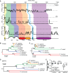
Recent Evolution of TYRO3, AXL, and MERTK. (A) Identification of regions of positive and purifying selection by Ka/Ks analysis, averaged over ten neighboring amino acids. Ka/Ks > 1.5 indicate positive selection, Ka/Ks < 0.5 indicate purifying selection (horizontal dotted lines). Major structural domains are indicated by colored shading; a generalized domain diagram is illustrated at the bottom of the figure. SP = signal peptide, Ig = immunoglobulin domain, Fbg = fibrinogen-like domain, TM = transmembrane domain, Kinase = tyrosine kinase domain. (BD) Maximum-likelihood evolutionary trees of TYRO3 (B), AXL (C), and MERTK (D) containing representative members of the hominini (orange), the apes (hominini plus green), old-world monkeys (blue) and new-world monkeys (red). Scales indicate the degree of evolutionary divergence, numbers at branch-points indicate bootstrap values. (E) Pairwise distances (Z-score) comparing the estimated number of synonymous substitutions per synonymous site (dS) and the number of nonsynonymous substitutions per nonsynonymous site (dN) between the human and primate MERTK grouped into clades containing all primate, all apes, and hominids. * = P < 0.05, n.s. = P > 0.05 compared with the primate-ancestral gene, 2-way ANOVA with Tukey Correction.
F<sc>ig</sc>. 2.
Fig. 2.
Reconstruction of Recent Evolution in the MERTK Signal Peptide and Transmembrane Domain. (A and B) Alignments of the human, hominid-ancestral, and primate-ancestral signal peptide DNA (A) and protein (B) sequence. The signal peptide cleavage point is indicated by the vertical arrow. (C) Predicted non-B DNA structure and SNP’s in the MERTK signal peptide. Tandem GCT repeats are indicated by horizontal arrows, the signal peptide cleavage point is indicated by the vertical arrow. (DE) DNA (D) and protein (E) alignments of the human, hominid-ancestral and primate-ancestral TMD, and membrane-proximal regions. Horizontal line indicates the TMD. Ancestral sequences were inferred using a maximum-likelihood approach.
F<sc>ig</sc>. 3.
Fig. 3.
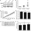
Influence of MERTK Signal Peptide Evolution on Protein Processing and Expression Level. MERTK bearing a human, ancestral-hominid, or ancestral-primate signal peptide were expressed in HeLa cells and their secretion, total expression, and half-lives quantified. (A and B) Total MERTK expression as quantified by the ratio of MERTK expression to the expression of a transfection control under the control of the same promotor (CD93). (A) Representative immunoblot, (B) quantification of MERTK expression, normalized to CD93. (C and D) Secretion of de novo synthesized MERTK was quantified by saturation-labeling of cell-surface HA-MERTK-GFP with Cy3, followed by quantification of the GFP:Cy3 ratio with fluorescence microscopy, (C) change in GFP:Cy3 ratio, normalized to time 0, and (D) MERTK export rate, quantified as the slope of panel A. (E and F) Quantification of MERTK half-life, (E) representative immunoblot, and (F) calculated half-life. n = 5–10. n.s. = P > 0.05, * = P < 0.05, compared with Human, Kruskal–Wallis test with Dunn correction.
F<sc>ig</sc>. 4.
Fig. 4.
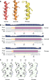
Protein Modeling of MERTK Transmembrane Domain. Amino acids 506–533 of human MERTK and the equivalent portion of the ancestral-hominid and ancestral-primate MERTK sequences, containing the TMD plus 10 N-terminal and 10 C-terminal residues were subjected to protein structure predictive modeling. (A) Ribbon diagram of the predicted structure and length of the MERTK TMD. (B) Confidence of helical structure versus nonhelical structure prediction. (C) Hydrophobic cluster analysis projected onto a 2D model of the MERTK TMD. Hydrophobic clusters appear in boxes, nonhydrophobic residues are in diamonds.
F<sc>ig</sc>. 5.
Fig. 5.
Evolution in MERTK Clustering and Avidity Driven by Transmembrane Domain Evolution. HeLa cells transfected with constructs containing human MERTK-GFP modified to contain the hominid or ape ancestral TMD were quantified for MERTK self-clustering, avidity, and enveloped viral binding. (A) Total Internal Reflection Microscopy (TIRF) and super-resolution ground state microscopy images (GSDM) of MERTK TMD constructs in the basolateral membrane. Boxes in the TIRF image indicate the 5 × 5 µm2 region shown in the GSDM image, scale bars are 1 µm. (B) Quantification of MERTK clustering by the radial distribution function G(r). (C) Clustering of MERTK TMD constructs, expressed as the area under the G(r) curve ± SEM, normalized to the degree of clustering observed in the primate-ancestral construct. (D) MERTK cluster radius, plotted as median ± interquartile range (whiskers = 5th/95th percentile). (E) Binding of apoptotic cell mimics by MERTK bearing human, hominid-ancestral or primate-ancestral TMDs. (F) Binding of apoptotic cell mimics by MERTK bearing human, hominid-ancestral or primate-ancestral TMDs at increasing shear stress. (G) Correlation between low-, medium-, and high-MERTK expression levels and the shear stress at which 50% of apoptotic cell mimics were retained on transfected cells. (H) Binding of Ebola VLPs to untransfected cells (Ctrl) or to cells expressing MERTK bearing Human (Hu), Hominid-ancestral (Ho) or Primate-ancestral (Pm) signal peptides, and TMDs. n = 3; * = P < 0.05 compared with Human, † compared with Hominid, n.s. = P > 0.05, ANOVA with Tukey correction.

Similar articles

Cited by

References

    1. 1000 Genomes Project Consortium, Auton A, Brooks LD, Durbin RM, Garrison EP, Kang HM, Korbel JO, Marchini JL, McCarthy S, McVean GA, et al.2015. A global reference for human genetic variation. Nature 526:68–74. - PMC - PubMed
    1. Bakewell M. a, Shi P, Zhang J.. 2007. More genes underwent positive selection in chimpanzee evolution than in human evolution. Proc Natl Acad Sci U S A. 104:7489–7494. - PMC - PubMed
    1. Bansal M, Kumar A, Yella VR.. 2014. Role of DNA sequence based structural features of promoters in transcription initiation and gene expression. Curr Opin Struct Biol. 25:77–85. - PubMed
    1. Best SM. 2013. Viruses play dead to TAMe interferon responses. Cell Host Microbe 14:117–118. - PMC - PubMed
    1. Bhattacharyya S, Zagórska A, Lew ED, Shrestha B, Rothlin CV, Naughton J, Diamond MS, Lemke G, Young JAT.. 2013. Enveloped viruses disable innate immune responses in dendritic cells by direct activation of TAM receptors. Cell Host Microbe 14:136–147. - PMC - PubMed

Publication types

MeSH terms