. 2017 Jun 1;9(3):220-230.
doi: 10.1093/jmcb/mjx010.

Human apo-SRP72 and SRP68/72 complex structures reveal the molecular basis of protein translocation

Affiliations

Human apo-SRP72 and SRP68/72 complex structures reveal the molecular basis of protein translocation

Yina Gao et al. J Mol Cell Biol. .

Abstract

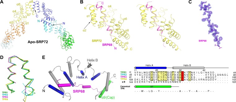
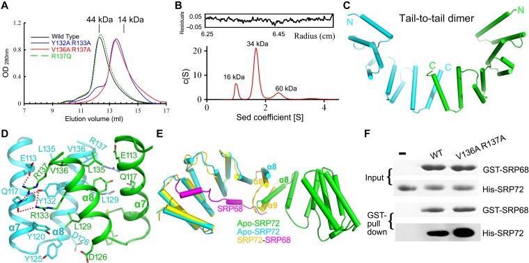
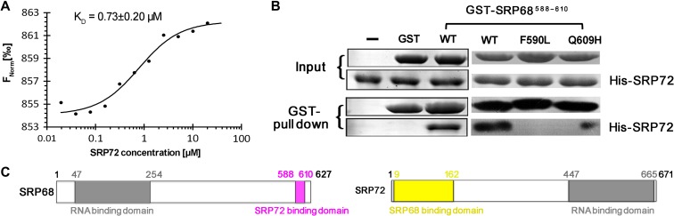
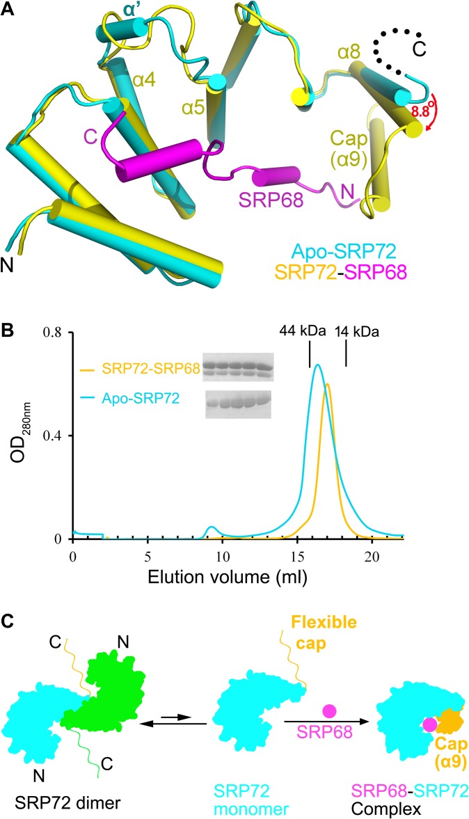
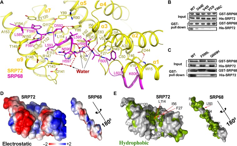
The co-translational targeting or insertion of secretory and membrane proteins into the endoplasmic reticulum (ER) is a key biological process mediated by the signal recognition particle (SRP). In eukaryotes, the SRP68-SRP72 (SRP68/72) heterodimer plays an essential role in protein translocation. However, structural information on the two largest SRP proteins, SRP68 and SRP72, is limited, especially regarding their interaction. Herein, we report the first crystal structures of human apo-SRP72 and the SRP68/72 complex at 2.91Å and 1.7Å resolution, respectively. The SRP68-binding domain of SRP72 contains four atypical tetratricopeptide repeats (TPR) and a flexible C-terminal cap. Apo-SRP72 exists mainly as dimers in solution. To bind to SRP68, the SRP72 homodimer disassociates, and the indispensable C-terminal cap undergoes a pronounced conformational change to assist formation of the SRP68/72 heterodimer. A 23-residue polypeptide of SRP68 is sufficient for tight binding to SRP72 through its unusually hydrophobic and extended surface. Structural, biophysical, and mutagenesis analyses revealed that cancer-associated mutations disrupt the SRP68-SRP72 interaction and their co-localization with ER in mammalian cells. The results highlight the essential role of the SRP68-SRP72 interaction in SRP-mediated protein translocation and provide a structural basis for disease diagnosis, pathophysiology, and drug design.

Keywords: SRP68; SRP72; cancer; crystal structures; protein translocation; protein–protein interaction; signal recognition particle.


Figures

Figure 1
Figure 1
Crystal structures of human apo-SRP72 and the SRP68/72 complex. (A) Overall structure of apo-SRP72. Six protomers are shown in different colors. (B) Stereoview of the SRP68/72 complex. SRP72 and SRP68 are colored yellow and magenta, respectively. (C) The composite simulated-annealing Fo-Fc ‘omit’ electron density map of SRP68 in the complex, contoured at 3.0 σ. (D) Structural overlay of the four TPRs in the SRP68/72 complex shows an atypical TPR and a same packing angle of the two helices. (E) Structure-based sequence alignment of all four TPRs in human SRP72 reveals inconsistence with canonical TPR motif. The canonical TPR sequence is shown at the bottom, and the conserved residues of SRP72 are shown in boxes.
Figure 2
Figure 2
Apo-SRP72 mainly exists as a dimer in solution. (A) SEC of SRP72 on a Superdex 75 10/300 GL column. The SEC profiles of the wild-type, Y132A/R133A, V136A/R137A double-mutants, and R137Q single mutant are colored black, blue, red, and green, respectively. Peak positions for two standard proteins are indicated as black lines on the top. (B) Analytical ultracentrifugation of the wild-type apo-SRP72. The c (S) distribution from SV analysis is shown. (C) Schematic representation of the apo-SRP72 as a tail-to-tail homodimer. The two molecules are colored cyan and green. The N- and C-termini of each monomer are labeled. (D) The interaction details of the tail-to-tail homodimer. The dimerization interface has two symmetric interaction networks, formed between two TPR4. Note that only one interaction interface is shown. (E) Structural overlay of SRP72 in the SRP68/72 complex (yellow) and in the tail-to-tail homodimer of apo-SRP72 (cyan and green). (F) The SRP72 monomeric mutant greatly increases the binding of GST-tagged SRP68 to SRP72.
Figure 3
Figure 3
The minimum fragment of SRP68 required to bind to SRP72. (A) MST measurements of the binding affinity of SRP68 for SRP72. The resulting binding curve from plotting the FNorm (‰) versus concentration was fit using a hyperbolic function to yield a KD of 0.73 ± 0.20 μM. (B) Residues 588−610 of SRP68 is enough for SRP72 binding. (C) Localization of the interaction regions in SRP68 and SRP72. A schematic diagram shows that both SRP68 and SRP72 consist of a RBD and a SRP72- or SRP68-binding domain.
Figure 4
Figure 4
SRP72 undergoes conformational changes upon binding to SRP68. (A) Structural comparison of apo-SRP72 (cyan) and the SRP72 (yellow)-SRP68 (magenta) complex. The α8 of the TPR4 rotated 8.8° towards (red arrow) to move closer to SRP68 when apo-SRP72 and SRP68-bound SRP72 were superimposed. (B) SEC profile of the SRP72 in the absence (cyan) or presence (yellow) of SRP68 (Superdex 200 10/300 GL column). Elution volumes of the molecular mass standards are marked at the top of the panel. The factions of the peaks were detected. The peak widths are significantly reduced, and the peaks become sharper after SRP68 binding. (C) SRP68–SRP72 binding model. A characteristic change occurred after SRP68 is bound to the concave surface of SRP72. The two molecules of apo-SRP72 are colored cyan and green. SRP68 and the cap are shown in magenta and orange, respectively.
Figure 5
Figure 5
The extensive interactions between SRP72 and SRP68. (A) Detailed representation of interactions between SRP72 (yellow) and SRP68 (magenta). Interacting residues are shown sticks. Water molecules involved in binding are represented as red spheres. (B and C) As shown by GST pull-down assays, cancer-associated mutations in SRP72 and SRP68 impair the interaction between SRP68 and SRP72. (D) The electrostatic potential (±2kBT) of the binding interface. The surface potential is displayed as a color gradient ranging from red (negative) to blue (positive). Note that SRP68 is rotated 180° around the axis to show the interface. (E) A surface representation of the binding interface, with hydrophobic residues in green. Residues contributing to the hydrophobic groove are labeled.
Figure 6
Figure 6
Mutations of key residues in SRP72–SRP68 interface impair the binding and localization. (A) Cellular co-localization between the ER and wild-type (WT) or mutants. Fixed cell images were obtained by using HeLa cells. SRP68 F590L mutation and SRP72 V53I, Y86C mutations showed diminished co-localization with ER compared to wild-type SRP68 and SRP72, respectively. Experiments were performed three times independently with similar results. Scale bar, 10 μm. (B) Binding abilities of the SRP72 and SRP68 mutants. Flag-tagged WT SRP68 or its mutants were from HCT116 cell lysate.

Similar articles

Cited by

References

    1. Akopian D., Shen K., Zhang X., et al. . (2013). Signal recognition particle: an essential protein-targeting machine. Annu. Rev. Biochem. 82, 693–721. - PMC - PubMed
    1. Andersen E.S., Rosenblad M.A., Larsen N., et al. . (2006). The tmRDB and SRPDB resources. Nucleic Acids Res. 34, D163–D168. - PMC - PubMed
    1. Becker M.M., Lapouge K., Segnitz B., et al. . (2017). Structures of human SRP72 complexes provide insights into SRP RNA remodeling and ribosome interaction. Nucleic Acids Res. 45, 470–481. - PMC - PubMed
    1. Buskiewicz I., Deuerling E., Gu S.Q., et al. . (2004). Trigger factor binds to ribosome-signal-recognition particle (SRP) complexes and is excluded by binding of the SRP receptor. Proc. Natl Acad. Sci. USA 101, 7902–7906. - PMC - PubMed
    1. Cingolani G., Petosa C., Weis K., et al. . (1999). Structure of importin-β bound to the IBB domain of importin-α. Nature 399, 221–229. - PubMed

Publication types