Comparative Study
. 2017 Jun 2;45(10):6217-6227.
doi: 10.1093/nar/gkx222.

Structure of acid deoxyribonuclease

Affiliations
Comparative Study

Structure of acid deoxyribonuclease

Armando Varela-Ramirez et al. Nucleic Acids Res. .

Abstract

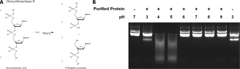
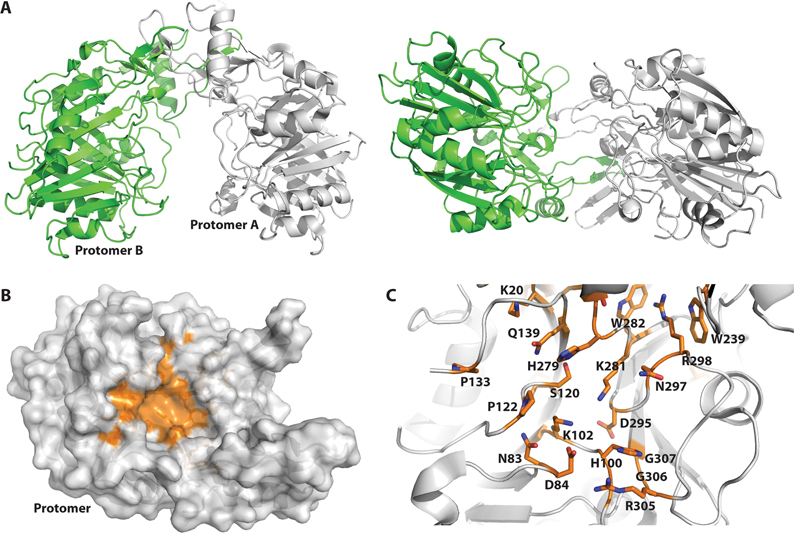
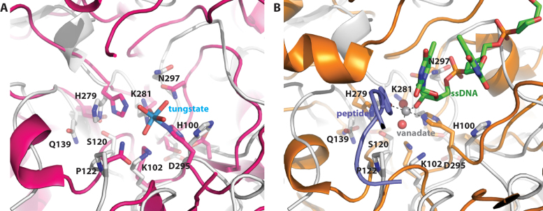
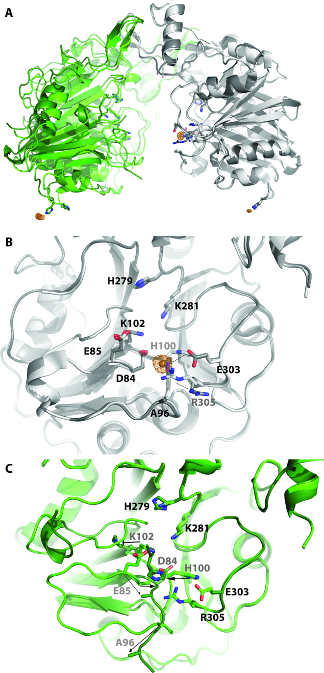
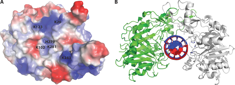
Deoxyribonuclease II (DNase II) is also known as acid deoxyribonuclease because it has optimal activity at the low pH environment of lysosomes where it is typically found in higher eukaryotes. Interestingly, DNase II has also been identified in a few genera of bacteria and is believed to have arisen via horizontal transfer. Here, we demonstrate that recombinant Burkholderia thailandensis DNase II is highly active at low pH in the absence of divalent metal ions, similar to eukaryotic DNase II. The crystal structure of B. thailandensis DNase II shows a dimeric quaternary structure which appears capable of binding double-stranded DNA. Each monomer of B. thailandensis DNase II exhibits a similar overall fold as phospholipase D (PLD), phosphatidylserine synthase (PSS) and tyrosyl-DNA phosphodiesterase (TDP), and conserved catalytic residues imply a similar mechanism. The structural and biochemical data presented here provide insights into the atomic structure and catalytic mechanism of DNase II.


Figures

Figure 1.
Figure 1.
Endonuclease activity of DNase II. (A) Deoxyribonuclease II (DNase II) hydrolyzes DNA in a metal ion-independent fashion at low pH to yield 3΄-phosphorylated and 5΄-hydroxyl nucleotides. (B) Cleavage of plasmid DNA by purified recombinant B. thailandensis DNase II (BthDNase II) under a variety of different pH conditions. Each cleavage buffer contained 10 mM EDTA as a chelating agent to remove divalent cations. In the absence of BthDNase II no cleavage is observed at neutral pH (lane 1) or at pH 3 (lane 9), although the lower pH results in an extra band consistent with single stranded plasmid. Smeared DNA at pH 4–5 represents BthDNase II-mediated plasmid degradation which is not observed at pH 3 and 6–9.
Figure 2.
Figure 2.
Crystal structure of B. thailandensis DNase II solved at 1.65 Å resolution. (A) Protomer A of the homodimeric BthDNase II structure is shown in gray ribbons and protomer B is shown in green ribbons either from the top at left or shown rotated 90° and viewed from the side at right. (B) Conserved residues are mapped onto the surface of the BthDNase II crystal structure and shown in orange (see supplemental Figure S1 for multiple sequence alignment). For simplicity, a single monomer of BthDNase II is shown viewed from the interior of the U-shaped clamp architecture. (C) Close-up of the active site, rotated slightly and zoomed in relative to panel B. Residues which are conserved across DNase II sequences are shown in sticks for their side chains and colored in CPK with orange carbon atoms. H100 and K102 comprise the first HxK catalytic motif whereas H279 and K281 comprise the second HxK catalytic motif.
Figure 3.
Figure 3.
Comparison of B. thailandensis DNase II and phospholipase D family members. (A) Overlay of BthDNase II (gray ribbons and sticks representation) with phospholipase D from Streptomyces in complex with tungstate (magenta ribbons and sticks rendering with tungsten shown in cyan and inner sphere coordinated waters in red). (B) Overlay of BthDNase II with a ternary complex of human TDP1 (orange ribbons and sticks rendering), DNA (green carbon CPK sticks), vanadate (dark gray and red spheres) and peptide.
Figure 4.
Figure 4.
Binding of Cu2+ to BthDNase II. (A) Overlay of the 1.65 Å resolution apo data set (chain A in gray, chain B in green) with the 1.75 Å resolution Cu2+ bound data set (chain A in dark gray, chain B in dark green, Cu2+ shown as copper colored spheres). Alignment was done via chain A. Anomalous maps are shown as copper colored mesh contoured at 4σ. (B) Active site of chain A showing movement of H100 upon binding Cu2+ along with movement of R305. Residues which move are labeled in gray and movement is shown with arrows. (C) Active site of chain B (via alignment of chain B) showing several loops which become disordered or change their conformation upon Cu2+ binding to H100 of chain A. Residues which move are labeled in gray and movement is shown with arrows.
Figure 5.
Figure 5.
Hypothetical model for DNA recognition by BthDNase II. (A) Surface electrostatics of a single BthDNase II protomer viewed from the interior of the homodimeric U-shaped clamp-like structure. Catalytic residues K102, H279, and H281 are visible from this vantage point, whereas H100 is not. Additional positively charged residues which line the central cavity K20, K123 and K243 are labeled. (B) Hypothetical model for DNA recognition by BthDNase II generated via manual docking of an ideal B-form DNA into the central cavity of the BthDNase II homodimer.

References

    1. Evans C.J., Aguilera R.J.. DNase II: genes, enzymes and function. Gene. 2003; 322:1–15. - PubMed
    1. Yang W. Nucleases: diversity of structure, function and mechanism. Q. Rev. Biophys. 2011; 44:1–93. - PMC - PubMed
    1. Steitz T.A. A mechanism for all polymerases. Nature. 1998; 391:231–232. - PubMed
    1. Shiokawa D., Tanuma S.. DLAD, a novel mammalian divalent cation-independent endonuclease with homology to DNase II. Nucleic Acids Res. 1999; 27:4083–4089. - PMC - PubMed
    1. Yamanaka M., Tsubota Y., Anai M., Ishimatsu K., Okumura M., Katsuki S., Takagi Y.. Purification and properties of acid deoxyribonucleases of human gastric mucosa and cervix uteri. J. Biol. Chem. 1974; 249:3884–3889. - PubMed

Publication types

MeSH terms