. 2017 Mar 1;68(7):1625-1637.
doi: 10.1093/jxb/erx034.

The complex character of photosynthesis in cucumber fruit

Affiliations

The complex character of photosynthesis in cucumber fruit

Xiaolei Sui et al. J Exp Bot. .

Abstract

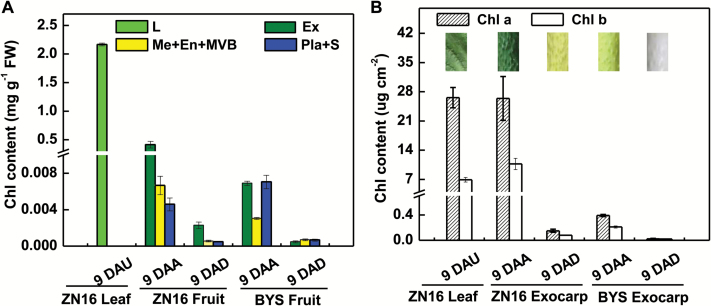
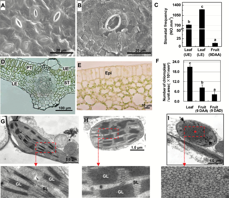
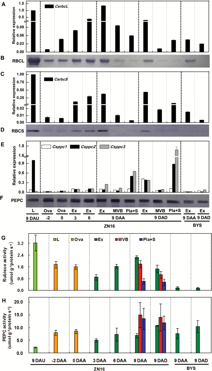
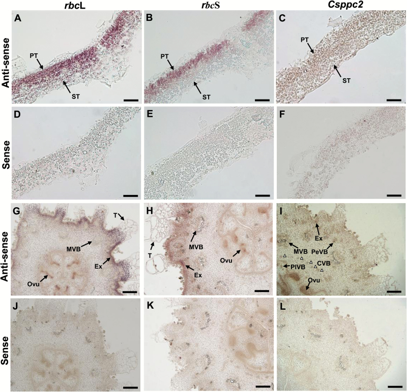
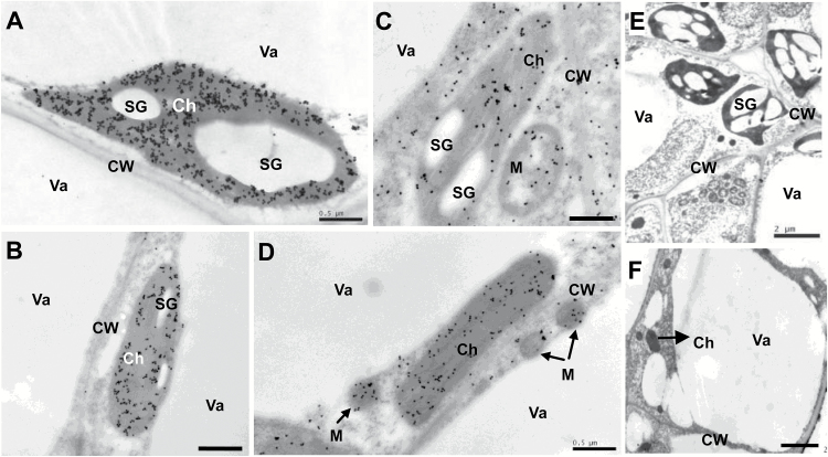
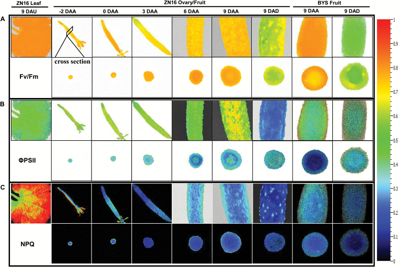
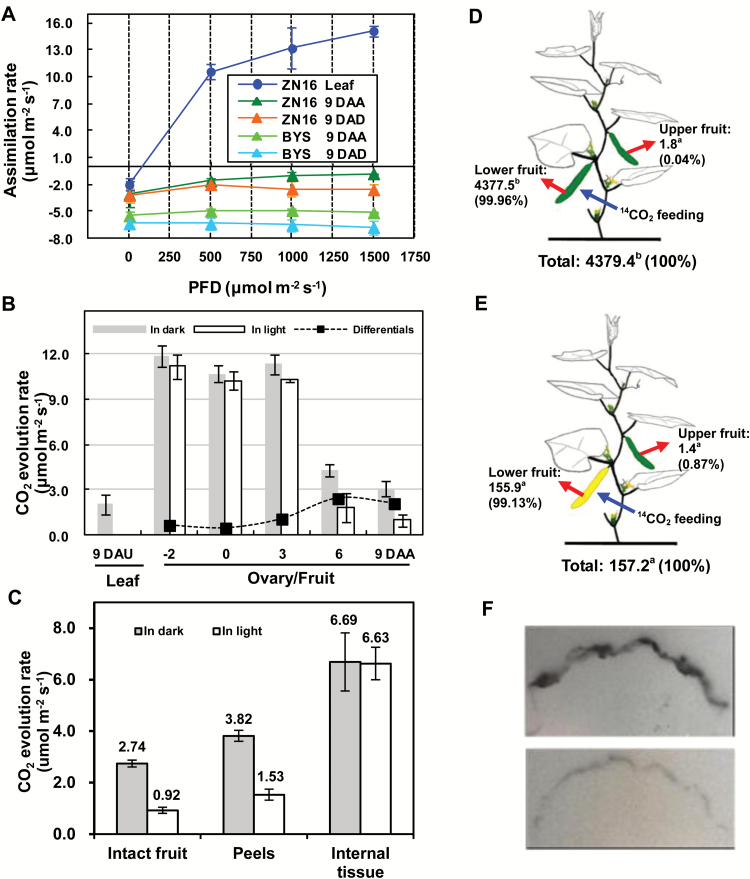
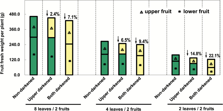
The surface area of a mature green cucumber (Cucumis sativa L.) fruit is comparable with that of a functional leaf, but the characteristics of fruit photosynthesis and its contribution to growth are poorly understood. Here, the photosynthetic properties of two genotypes of cucumber (dark green and light green fruits) were studied using a combination of electron microscopy, immunogold enzyme localization, chlorophyll fluorescence imaging, isotope tracer, and fruit darkening techniques. Chlorophyll content of the exocarp is similar to that of leaves, but there are no distinctive palisade and spongy tissues. The efficiency of PSII is similar to that in leaves, but with lower non-photochemical quenching (NPQ). Ribulose-1,5-bisphosphate carboxylase/oxygenase (Rubisco) is found mainly in the exocarp, while phosphoenolpyruvate carboxylase (PEPC) is primarily localized to vascular bundles and placenta tissue. Rubisco and PEPC expression at both transcriptional and translational levels increases concurrently during fruit growth. The contribution of fruit photosynthesis in exocarp to its own C accumulation is 9.4%, while ~88% of respiratory CO2 in fruit was captured and re-fixed. Photosynthesis by cucumber fruits, through direct fixation of atmospheric CO2 and recapture of respired CO2, as verified by 14CO2 uptake and gas exchange, makes an important contribution to fruit growth.

Keywords: Chloroplast; PEPC (phosphoenolpyruvate carboxylase); Rubisco (ribulose-1,5-bisphosphate carboxylase/oxygenase).; cucumber; fruit photosynthesis; respiration.


Figures

Fig. 1.
Fig. 1.
Chlorophyll content in fruits and leaves based on per unit fresh weight (A) and surface area (B). Images in (B) were obtained from a leaf at 9 DAU and a fruit at 9 DAA/9 DAD, respectively, illustrated by different colors. Error bars represent the SD, n=3. DAA, days after anthesis; DAD, days after darkening; DAU, days after unfolding; Chl, chlorophyll; En, endocarp; Ex, exocarp; L, leaf; Me, mesocarp; Pla, placenta; S, seeds; MVB, main vascular bundle.
Fig. 2.
Fig. 2.
Anatomical features of chloroplasts of cucumber exocarp. (A, B) Scanning electron micrographs of the lower epidermis of a mature leaf (9 DAU) (A) and epidermis of fruit (9 DAA) (B). The stomata are covered with cuticular wax on the fruit epidermis. (C) Stomatal frequency. (D, E) Cross-section of a leaf at 9 DAU (D) and a fruit at 9 DAA (E). (F) Number of chloroplasts per unit area. (G–I) Transmission electron micrographs of chloroplasts distributed in palisade cells of a mature leaf (9 DAU) (G), parenchyma cells of fruit (9 DAA) (H), and darkened fruit (9 DAB) (I), respectively. Variety: ‘ZN16’. Means followed by different letters indicate statistically significant differences according to Tukey’s test (P<0.05) [(n=50 in (C); n=10 in (F)]. Epi, epidermis; GL, grana lamellae; LE, lower epidermis; PT, palisade tissue; SL, stroma lamella; ST, spongy tissue; UE, upper epidermis.
Fig. 3.
Fig. 3.
Expression and activities of Rubisco and PEPC in cucumber fruits. (A–F) Quantitative real-time PCR analysis of rbcL (A), rbcS (C), and ppc (E) mRNA levels, and immunoblot analysis of the Rubisco large (RBCL) (B) and small (RBCS) (D) subunits and PEPC (F) using antibodies to each of the respective proteins. Experiments for the quantification of protein levels were repeated three times, yielding similar results. (G, H) Enzymatic activities of Rubisco (G) and PEPC (H), which were calculated per protein concentration. Error bars represent the SD, n=3. For abbreviations, see Fig. 1.
Fig. 4.
Fig. 4.
In situ hybridization of rbcL, rbcS, and ppc transcripts in cucumber fruits. (A–F) Leaf cross-sections (0–1 DAU) hybridized with the rbcL, rbcS, and Csppc2 antisense (A–C) and sense (D–F) probes, respectively. (G–L) Young ovary/fruit cross-sections (–2 –0 DAA) hybridized with the rbcL, rbcS, and Csppc2 antisense (G–I) and sense (J–L) probes, respectively. White triangles in (I) indicate the CVB. CVB, carpel vascular bundle; Ovu, ovule; PeVB, peripheral vascular bundle; PlVB, placenta vascular bundle; T, trichome; for other abbreviations, see Figs 1 and 2. Scale bars=50 µm in (A–F) and 200 µm in (G–L).
Fig. 5.
Fig. 5.
Immunogold localization of Rubisco and PEPC in cucumber fruits. (A, B) Rubisco (gold particles) was localized to chloroplasts in leaf (A) and ovary/fruit (B). (C, D) PEPC was targeted to the chloroplast, cytoplasm, and mitochondria in leaf (C) and ovary/fruit (D). (E, F) The controls of leaf (E) and ovary/fruit (F) without incubation with antibodies; no substantial signals were detected. Sections were prepared from fully expanded leaves and ovaries/fruits on the day of anthesis. Scale bars=0.5 µm in (A–D), and 2 µm in (E, F). Ch, chloroplast; CW, cell wall; M, mitochondrion; SG, starch grain; Va, vacuole.
Fig. 6.
Fig. 6.
Chlorophyll fluorescence imaging of cucumber fruits. (A–C) Fluorescence images correspond to Fv/Fm (A), ΦPSII (B), and NPQ (C). The upper row shows images of the whole or partial fruits; in the lower row are the corresponding cross-sections. All images were normalized to a false color bar (see right column). The pixel value display is based on a false-color scale ranging from blue (0.0), green, yellow, to red (ending at 1.0). The analyses of Fv/Fm were carried out on dark-adapted fruits and leaves, while those of ΦPSII and NPQ were at a light intensity of 500 μmol quanta m–2 s–1. Fv/Fm, maximum PSII quantum yield; ΦPSII, steady-state PSII quantum yield; NPQ, steady-state non-photochemical quenching.
Fig. 7.
Fig. 7.
CO2 assimilation and 14C feeding in cucumber fruits. (A) The response curve of assimilation rate to photon flux density (PFD). (B) CO2 evolution rate of exocarp (peels) in the dark and illuminated (1000 µmol quanta m–2 s–1 irradiance) conditions during ‘ZN16’ fruit development. The net photosynthetic rate per unit fruit surface area (black squares) was the difference between the CO2 evolution rates in the light and dark. All data above were determined at ambient CO2 between 390 mbar and 410 mbar and at air temperature of 28°C. (C) CO2 evolution rate of intact fruit, peels, and internal tissue under dark and light (1000 µmol quanta m–2 s–1 irradiance) conditions, respectively (Variety ‘ZN16’, 9 DAA). Error bars represent the SD, n=3. (D, E) Allocation of 14C in cucumber fruits. Lower non-bagged (D) or bagged (E) fruits were fed with 3.7 × 10⁶ Bq 14CO2 (blue arrows). After 24 h, specific activities (Bq g–1 DW) of lower and upper fruits (red arrows) were determined. The means (n=3) followed by different letters indicate statistically significant differences according to Tukey’s test (P<0.05). Values in parentheses are percentages of the total measurements. (F) 14C autoradiograph of non-darkened fruit (upper photo) and darkened fruit (lower photo) at 1 h after the ending of 14CO2 feeding.
Fig. 8.
Fig. 8.
Effect of darkening cucumber fruits on fruit yield. For treatment of bagged fruits, see Supplementary Fig. S1. Values represent the average of 20 replicates. Values on the bars are the percentage decrease of fresh fruit weight after 9 d dark treatment. Green and yellow bars indicate non-darkened and darkened fruits, respectively.

Similar articles

Cited by

References

    1. Araújo WL, Nunes-Nesi A, Osorio S, et al. 2011. Antisense inhibition of the iron–sulphur subunit of succinate dehydrogenase enhances photosynthesis and growth in tomato via an organic acid-mediated effect on stomatal aperture. The Plant Cell 23, 600–627. - PMC - PubMed
    1. Aschan G, Pfanz H. 2003. Non-foliar photosynthesis—a strategy of additional carbon acquisition. Flora 198, 81–97.
    1. Baker NR. 2008. Chlorophyll fluorescence: a probe of photosynthesis in vivo. Annual Review of Plant Biology 59, 89–113. - PubMed
    1. Blanke MM, Lenz F. 1989. Fruit photosynthesis. Plant, Cell and Environment 12, 31–46.
    1. Bradford MM. 1976. A rapid and sensitive method for the quantitation of microgram quantities of protein utilizing the principle of protein–dye binding. Analytical Biochemistry 72, 248–254. - PubMed

Publication types

MeSH terms

Substances