. 2017 Mar 1;68(7):1441-1450.
doi: 10.1093/jxb/erx047.

NETWORKED 3B: a novel protein in the actin cytoskeleton-endoplasmic reticulum interaction

Affiliations

NETWORKED 3B: a novel protein in the actin cytoskeleton-endoplasmic reticulum interaction

Pengwei Wang et al. J Exp Bot. .

Abstract

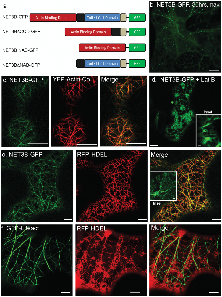
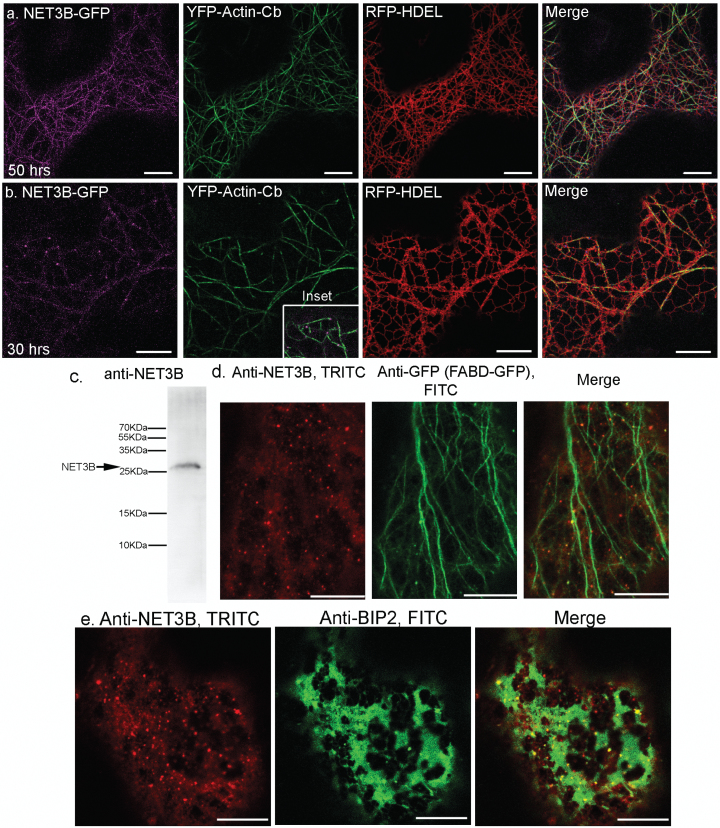
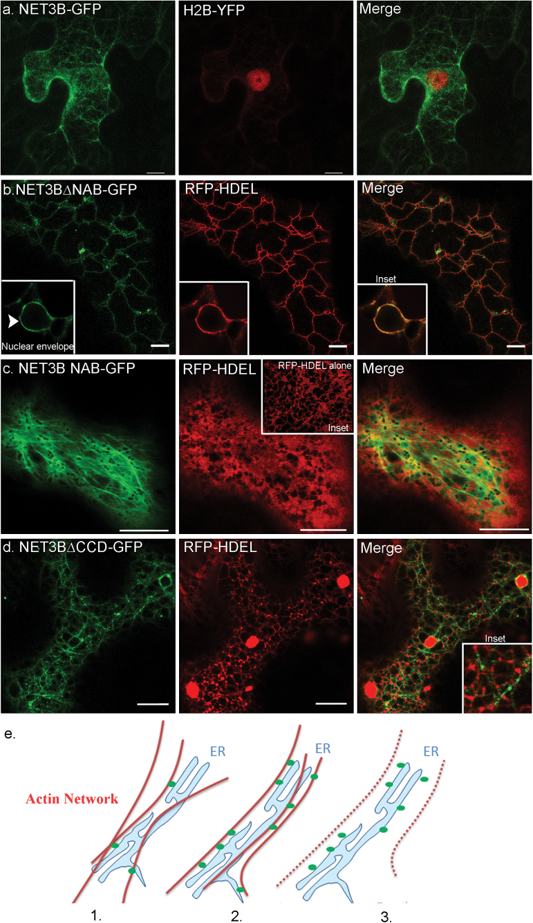
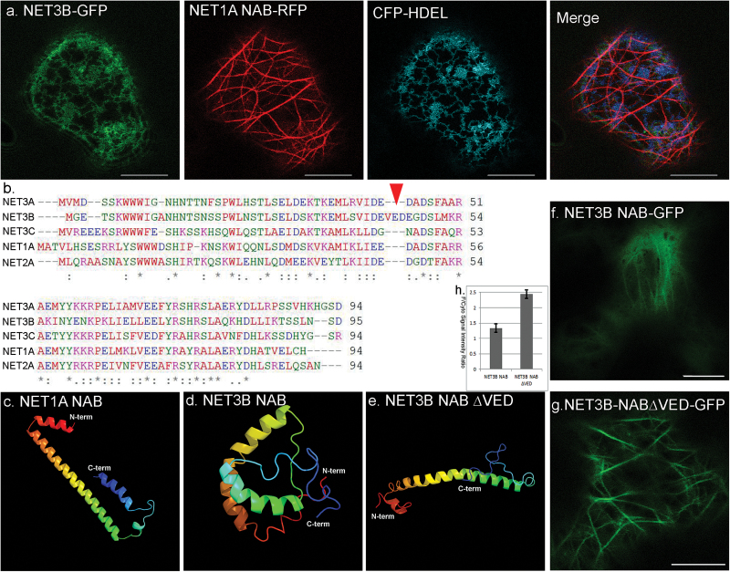
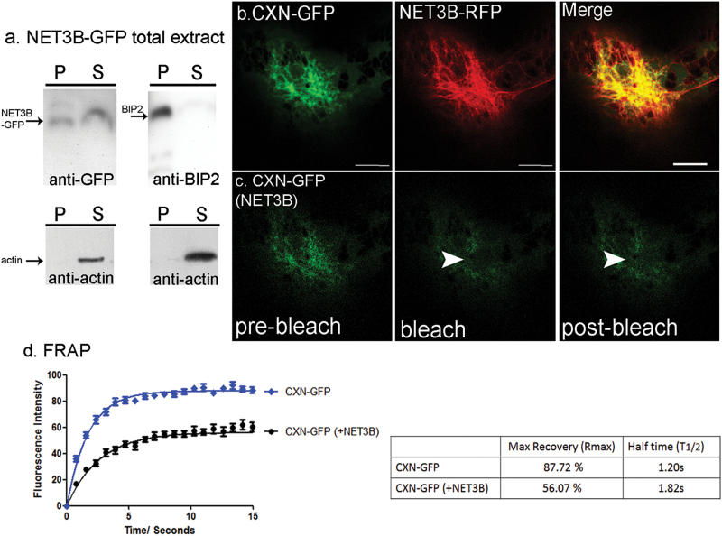
In plants movement of the endoplasmic reticulum (ER) is dependent on the actin cytoskeleton. However little is known about proteins that link the ER membrane and the actin cytoskeleton. Here we identified a novel protein, NETWORKED 3B (NET3B), which is associated with the ER and actin cytoskeleton in vivo. NET3B belongs to a superfamily of plant specific actin binding proteins, the NETWORKED family. NET3B associates with the actin cytoskeleton in vivo through an N-terminal NET actin binding (NAB) domain, which has been well-characterized in other members of the NET family. A three amino acid insertion, Val-Glu-Asp, in the NAB domain of NET3B appears to lower its ability to localize to the actin cytoskeleton compared with NET1A, the founding member of the NET family. The C-terminal domain of NET3B links the protein to the ER. Overexpression of NET3B enhanced the association between the ER and the actin cytoskeleton, and the extent of this association was dependent on the amount of NET3B available. Another effect of NET3B overexpression was a reduction in ER membrane diffusion. In conclusion, our results revealed that NET3B modulates ER and actin cytoskeleton interactions in higher plants.

Keywords: Actin cytoskeleton; N. benthamiana.; NET superfamily; endomembrane system; endoplasmic reticulum.


Figures

Fig. 1.
Fig. 1.
NET3B-GFP co-localizes with both the actin cytoskeleton and the ER in transformed N. benthamiana leaf epidermal cells. N. benthamiana leaf epidermal cells transiently transfected with fluorescent protein constructs either singly or in combination with other construct(s) as shown in each panel. (a) Graphical illustration of the construction of NET3B fusions. (b) 3D maximum projection of NET3B-GFP expressing cells with low expression. NET3B-GFP labels numerous actin cytoskeleton-associated puncta, producing the typical ‘beads-on-a-string’ pattern characteristic of NET family proteins. (c) NET3B-GFP/YFP-actin-Cb expressing cells. NET3B-GFP localizes to filamentous structures that are also labelled with the actin marker. (d) NET3B-GFP redistributes to the ER membrane when the actin cytoskeleton is depolymerised by latrunculin B. The inset picture shows that GFP-Lifeact becomes cytoplasmic after latrunculin B treatment. (e) NET3B-GFP/RFP-HDEL expressing cells. The expression of NET3B-GFP enhances the association between the ER and the actin cytoskeleton. Consequently, the morphology of the RFP-HDEL labelled ER network shows greater alignment with the actin network. The inset picture shows that full length NET3B without a tag is also able to enhance the association between the actin cytoskeleton and the ER. (f) GFP-Lifeact/RFP-HDEL expressing cells. The actin and ER networks exhibit little alignment with each other in contrast to (e). Lat B, latrunculin B. Scale bar, 10 µm.
Fig. 2.
Fig. 2.
Endogenous Arabidopsis NET3B co-localize with the actin and ER networks. (a–b) NET3B-GFP (magenta) co-expressed with YFP-actin-Cb (green) and RFP-HDEL (red) at different time points after infiltration into N. benthamiana leaves. The same detection settings were used for imaging GFP. Strong alignment between the ER and actin cytoskeleton was seen in (a) but not in (b). The NET3B puncta seen in (b) align with the actin cytoskeleton (inset) and are associated with the ER membrane. (c) Western blot of total protein extract from Arabidopsis seedlings probed with a polyclonal NET3B antibody (anti-NET3B) showing a clear band at around 25 kDa. (d) Immunofluorescence images of cotyledon cells of an Arabidopsis line stably expressing FABD2-GFP, which marks actin. Cells stained with anti-NET3B (TRITC, red) and anti-GFP antibodies (FITC, green). Endogenous NET3B was detected in both the cytoplasm and as punctate structures, which aligned with the actin network. (e) Immunofluorescence images showing that the anti-NET3B labelled puncta also associated with the ER network, which is labelled with anti-BIP2 (FITC, green). Scale bar, 10 µm.
Fig. 3.
Fig. 3.
NET3B co-localizes with the actin and ER networks through its N-terminal NAB domain and a C-terminal ER binding domain, respectively. Domain deletion mutants of NET3B were fused in frame to GFP and transiently expressed with RFP-HDEL in N. benthamiana leaf epidermal cells. (a) 3D projection of cells expressing NET3B-GFP and the nuclear marker H2B-YFP. Note: unlike most ER membrane proteins, NET3B-RFP does not label the nuclear envelope. (b) NET3B∆NAB-GFP/RFP-HDEL expressing cells. NET3B∆NAB localizes to the ER network and the nuclear envelope (inset). (c) NET3B-NAB-GFP expressing cells. NET3B-NAB-GFP labels the actin cytoskeleton, with a strong cytoplasmic background. Under these conditions, the ER network becomes more cisternae-like when compared with cells expressing RFP-HDEL (inset). (d) NET3B∆CCD-GFP expressing cells. NET3B∆CCD-GFP labels the actin cytoskeleton, with numerous puncta aligned along the actin network. The ER network is disrupted and its association with the NET3B∆CCD-GFP labeled actin cytoskeleton is much reduced. (e) Proposed model of NET3B interaction with the ER. (1) NET3B associates with the ER and the actin cytoskeleton at specific membrane foci. (2) NET3B overexpression brings the actin cytoskeleton and the ER network closer together by increasing the number of sites of interaction. (3) NET3B localizes to the ER when the actin cytoskeleton depolymerises. Scale bar, 10 µm.
Fig. 4.
Fig. 4.
NET3B has an insertion in its NAB domain that reduces its ability to associate with the actin cytoskeleton in vivo. (a) N. benthamiana leaf epidermal cells expressing NET3B-GFP/NET1A-NAB-RFP/CFP-HDEL. NET3B-GFP is not able to associate with the actin cytoskeleton in the presence of NET1A-NAB-RFP. Note: the actin cytoskeleton is only labelled with NET1A-NAB-RFP and NET3B-GFP localizss to the ER as determined by its co-localization with CFP-HDEL. (b) Sequence alignment of the NAB domain from NET1A, NET2A, NET3A, NET3B and NET3C. The three amino acid insertion, VED, is a unique feature of NET3B. (c–d) Predicted 3D structures of the NAB domains listed in (b) analysed using Phyre2. Significant differences are seen in the 3D structure of the NAB domains of NET1A NET3B. The arrow indicates the approximate position of the VED insertion of NET3B. (e) Once the VED motif is removed from the NAB domain of NET3B, its predicted 3D structure is very similar to NET1A. (f–g) N. benthamiana leaf epidermal cells expressing NET3B-NAB-GFP and NET3B-NAB∆VED -GFP where the VED motif is absent. A stronger cytoplasmic signal is seen in NET3B-NAB-GFP expressing cells. (h) Quantification of the ratio between the fluorescence intensities of the cytoplasm and the actin cytoskeleton in (f–g), further illustrating the stronger cytoplasmic signal seen in NET3B-NAB-GFP expressing cells. Scale bar, 10 µm.
Fig. 5.
Fig. 5.
NET3B-GFP overexpression restricts membrane diffusion. (a) Western blots of high-speed ultracentrifuged pellets and supernatants from N. benthamiana leaf extracts expressing NET3B-GFP. (i) Anti-GFP antibody shows that NET3B-GFP is detected in the pellet, which harbours the ER membrane microsomal fraction and in the supernatant. Anti-actin antibody shows that actin is only present in the supernatant; (ii) The ER protein BIP2 is found only in the pellet, as detected by anti-BIP2 antibody. Again, anti-actin antibody revels actin is only present in the supernatant. Note: NET3B-GFP has the characteristics of a peripheral membrane protein. (b-c) Representative images of photobleached ER membrane imaged for CXN-GFP in the presence of NET3B-RFP. (d) FRAP analysis of CXN-GFP in CXN-GFP/NET3B-RFP expressing cells compared with cells only expressing CXN-GFP. Note: Movement of the ER membrane, as depicted by the recovery of CXN-GFP fluorescence, is signifcantly retarded in the presence of NET3B-RFP. This is reflected by a prolonged T1/2 and reduced Rmax. Scale bar,10 µm.
Fig. 6.
Fig. 6.
NET3B mutants, net3b-1 and net3b-2, exhibit no significant effects on the ER network. (a) NET3B::GUS expression in stably transformed Arabidopsis lines. GUS staining was seen in seedlings (1), developing seeds (2–3), anther/pollen (4) and pollen tubes (5). (b) Illustration of the position of the T-DNA insertions in net3b-1 and net3b-2 lines. (c) Western blot of total protein extracts from Arabidopsis flowers of Col-0, net3b-1 and net3b-2, probed with anti-NET3B antibody. Note: NET3B is absent from net3b-2, which a knockout mutant, while net3b-1 shows significantly reduced NET3B expression. (d) The amido black stain indicates equal protein loading. (e) Both wild type and net3b-2 plants are transformed with RFP-HDEL. Hypocotyl epidermal cells are shown. Note: the ER organisation looks similar and no obvious defects can be observed. Scale bar, 10 µm.

Similar articles

Cited by

References

    1. Breeze E, Dzimitrowicz N, Kriechbaumer V, et al. 2016. A C-terminal amphipathic helix is necessary for the in vivo tubule-shaping function of a plant reticulon. Proceedings of the National Academy of Sciences, USA 113, 10902–10907. - PMC - PubMed
    1. Boevink P, Oparka K, Santa Cruz S, Martin B, Betteridge A, Hawes C. 1998. Stacks on tracks: the plant Golgi apparatus traffics on an actin/ER network. The Plant Journal 15, 441–447. - PubMed
    1. Chen J, Stefano G, Brandizzi F, Zheng H. 2011. Arabidopsis RHD3 mediates the generation of the tubular ER network and is required for Golgi distribution and motility in plant cells. Journal of Cell Science 124, 2241–2252. - PubMed
    1. Deeks MJ, Calcutt JR, Ingle EK, et al. 2012. A superfamily of actin-binding proteins at the actin-membrane nexus of higher plants. Current Biology 22, 1595–1600. - PubMed
    1. Griffing LR, Gao HT, Sparkes I. 2014. ER network dynamics are differentially controlled by myosins XI-K, XI-C, XI-E, XI-I, XI-1, and XI-2. Frontiers in Plant Science 5, 218. - PMC - PubMed

MeSH terms

Substances