. 2017 Jul 1;34(7):1654-1668.
doi: 10.1093/molbev/msx107.

The Influence of HIV on the Evolution of Mycobacterium tuberculosis

Affiliations

The Influence of HIV on the Evolution of Mycobacterium tuberculosis

Anastasia S Koch et al. Mol Biol Evol. .

Abstract

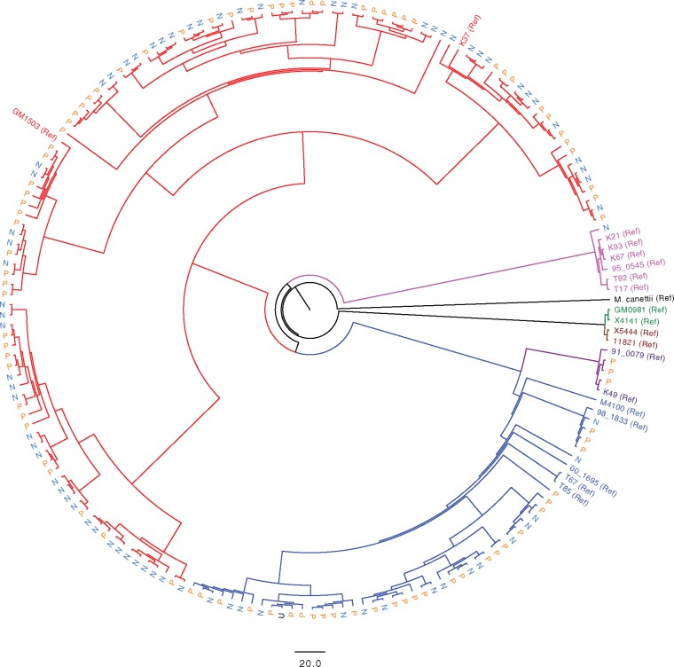
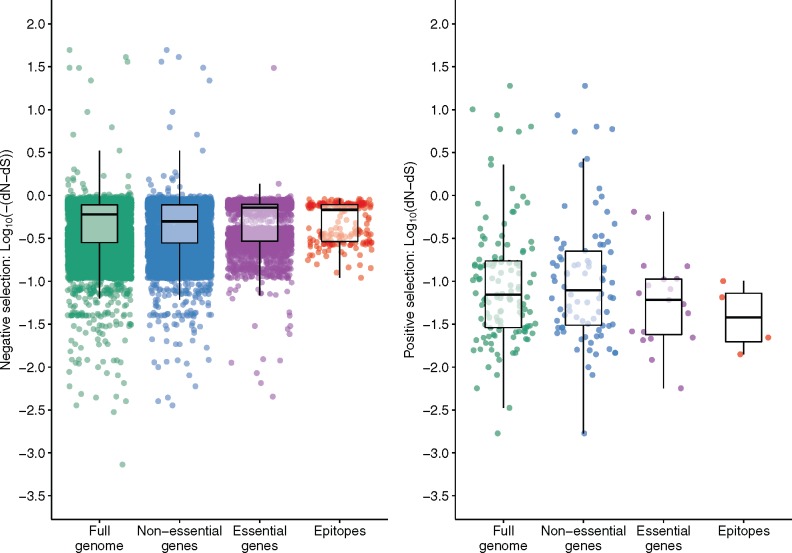
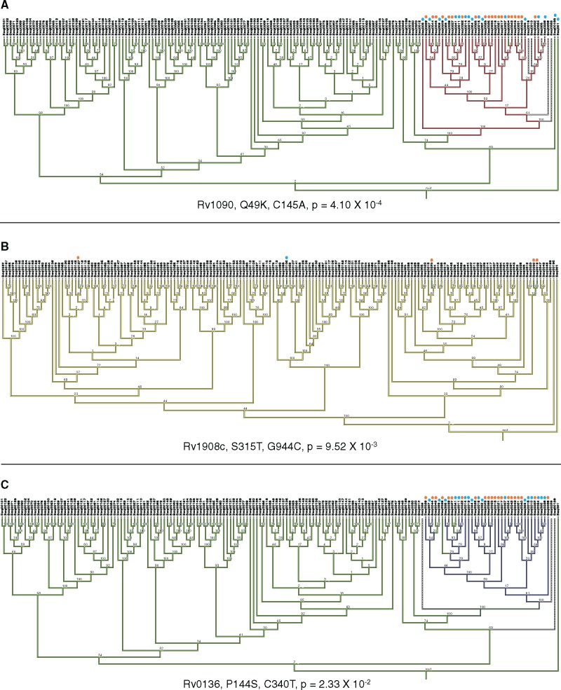
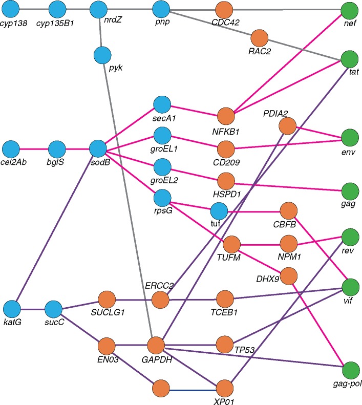
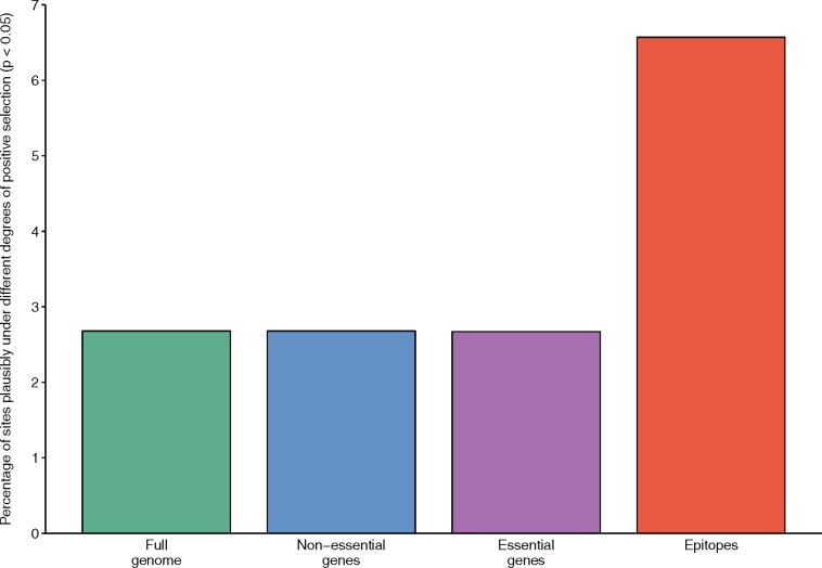
HIV significantly affects the immunological environment during tuberculosis coinfection, and therefore may influence the selective landscape upon which M. tuberculosis evolves. To test this hypothesis whole genome sequences were determined for 169 South African M. tuberculosis strains from HIV-1 coinfected and uninfected individuals and analyzed using two Bayesian codon-model based selection analysis approaches: FUBAR which was used to detect persistent positive and negative selection (selection respectively favoring and disfavoring nonsynonymous substitutions); and MEDS which was used to detect episodic directional selection specifically favoring nonsynonymous substitutions within HIV-1 infected individuals. Among the 25,251 polymorphic codon sites analyzed, FUBAR revealed that 189-fold more were detectably evolving under persistent negative selection than were evolving under persistent positive selection. Three specific codon sites within the genes celA2b, katG, and cyp138 were identified by MEDS as displaying significant evidence of evolving under directional selection influenced by HIV-1 coinfection. All three genes encode proteins that may indirectly interact with human proteins that, in turn, interact functionally with HIV proteins. Unexpectedly, epitope encoding regions were enriched for sites displaying weak evidence of directional selection influenced by HIV-1. Although the low degree of genetic diversity observed in our M. tuberculosis data set means that these results should be interpreted carefully, the effects of HIV-1 on epitope evolution in M. tuberculosis may have implications for the design of M. tuberculosis vaccines that are intended for use in populations with high HIV-1 infection rates.

Keywords: HIV coinfection; Mycobacterium tuberculosis; evolution; natural selection.


Figures

F<sc>ig</sc>. 1.
Fig. 1.
Phylogeny of M. tuberculosis strains isolated from HIV-1 coinfected and HIV-1 uninfected individuals. A maximum likelihood phylogenetic tree was constructed using RAxML (Stamatakis 2014). A reference set of strains (annotated as “Ref” on the tree) representative of the known global diversity of M. tuberculosis (Comas et al. 2010) was included to provide phylogenetic context for strains isolated in Khayelitsha, South Africa. M. canettii is included as an outgroup. Blue and red branches indicate strains belonging to lineage 2 and 4, respectively. Other lineage branches are coloured as in (Comas et al. 2010). Strains isolated from HIV-1 uninfected individuals (N) are indicated in light blue, and those from HIV-1 coinfected individuals (P) in orange. The scale bar indicates SNP differences. The tree with associated bootstrap values is available in the supporting information (supplementary fig. 1, Supplementary Material online).
F<sc>ig</sc>. 2.
Fig. 2.
Strengths of natural selection differ for different functional gene categories in M. tuberculosis. Estimates at 23,860 codon sites within the analyzed M. tuberculosis genomes were evaluated for nonsynonymous (dN) and synonymous substitution rates (dS) without considering HIV-1 status using the FUBAR method (Murrell et al. 2013). Analyzed codon sites were classified into essential, nonessential and epitope encoding regions (Sassetti et al. 2003). The left panel shows codons that are under negative selection (dN–dS < 0). Each dot represents the log10 of the −dN–dS values reported by the FUBAR analysis. The right panel shows codons under positive selection dN–dS>0, with each dot representing the log10 of dN–dS values. The boxplots represent the interquartile range (IQR), the median and the highest and lowest values within 1.5 times the IQR and outliers are captured by the scatterplot. Wilcoxon rank sum tests were performed by comparing the distribution of dN–dS values for all individual codons in each category: essential versus nonessential (P < 0.001); essential versus epitope (P = 0.192); nonessential versus epitope (P = 0.336).
F<sc>ig</sc>. 3
Fig. 3
Phylogenetic mapping of M. tuberculosis codon sites with highly significant P values for evidence of HIV-1 associated directional selection. MESQUITE (Maddison and Maddison, 2017) was applied to map the phylogenetic position of codon sites with intermediate to low MEDS associated P values. For each site the amino acid change, the nucleotide change and the degree of Bonferroni corrected P value support is indicated. M. tuberculosis strains with the mutation isolated from HIV-1 coinfected individuals are indicated with an orange dot, and those from HIV-1 uninfected individuals a blue dot. Phylogenetic trees were generated in RAxML (Stamatakis 2014) and bootstrap values from 1000 replicates are annotated on nodes. (A) The site with the strongest evidence of directional selection in HIV-1 coinfected individuals is shown. Branches that contain the ancestral cytosine are indicated in green, while those that contain adenine are shown in red, dashed branches indicated branches for which there was no call at that site for that strain. (B) Phylogenetic mapping of the S513T mutation in the katG gene (Rv1908c). This substitution involves a single nucleotide substitution from guanine (in yellow) to cytosine (in green). (C) The third codon site very strong evidence of HIV-1 associated directional selection occurs in cyp138 (Rv0136). The nucleotide change at this site is from cytosine (in green) to thymine (in purple).
F<sc>ig</sc>. 4.
Fig. 4.
Inferred functional interactions between M. tuberculosis genes with codons under HIV-1 associated directional selection and human and HIV-1 genes. A functional interaction analysis was conducted to investigate the biological significance of M. tuberculosis genes with evidence for directional selection influenced by HIV-1 coinfection. Data for intraspecies human–human, M. tuberculosis–M. tuberculosis interactions was retrieved from STRING (Szklarczyk et al. 2015) and REACTOME (Fabregat et al. 2016; Croft et al. 2014), with a cutoff confidence score of 0.7. Human–HIV-1 interactions were derived from the HIV-1 Human Protein Interaction Database (Fu et al. 2009) and human–M. tuberculosis interactions were retried from previously generated data (Rapanoel et al. 2013 and Huo et al. 2015). For both interspecies interactions, data was only included if previously reported in the literature. Blue circles represent genes from M. tuberculosis, orange circles from humans, and green circles from HIV-1.
F<sc>ig</sc>. 5.
Fig. 5.
Coding sites in epitope encoding regions are enriched for evidence of directional selection influenced by HIV-1 coinfection. Episodic directional selection in M. tuberculosis strains isolated from HIV-1 uninfected and HIV-1 coinfected individuals was evaluated using the MEDS method (Murrell et al. 2012). Sites that showed evidence of directional selection associated with phylogenetic branches separating HIV-1 uninfected and HIV-1 coinfected individuals (i.e., with an associated MEDS P value < 0.05, without Bonferroni multiple testing correction) were assigned to essential, nonessential and epitope encoding gene categories (Sassetti et al. 2003). The percentage of sites falling in each category is indicated by the bars. The site counts used to calculate these percentages can be seen in supplementary table 3, Supplementary Material online.

References

    1. Abraha A, Nankya IL, Gibson R, Demers K, Tebit DM, Johnston E, Katzenstein D, Siddiqui A, Herrera C, Fischetti L, et al.2009. CCR5- and CXCR4-tropic subtype C human immunodeficiency virus type 1 isolates have a lower level of pathogenic fitness than other dominant group M subtypes: implications for the epidemic. J Virol. 83:5592–5605. - PMC - PubMed
    1. Achtman M. 2008. Evolution, population structure, and phylogeography of genetically monomorphic bacterial pathogens. Annu Rev Microbiol. 62:53–70. - PubMed
    1. Achtman M. 2012. Insights from genomic comparisons of genetically monomorphic bacterial pathogens. Philos Trans R Soc Lond B Biol Sci. 367:860–867. - PMC - PubMed
    1. Anisimova M, Kosiol C.. 2009. Investigating protein-coding sequence evolution with probabilistic codon substitution models. Mol Biol Evol. 26:255–271. - PubMed
    1. Ariën KK, Vanham G, Arts EJ.. 2007. Is HIV-1 evolving to a less virulent form in humans?. Nat Rev Microbiol. 5:141–151. - PMC - PubMed

Publication types