. 2017 Mar 1;68(7):1613-1623.
doi: 10.1093/jxb/erx058.

Cell layer-specific patterns of cell division and cell expansion during fruit set and fruit growth in tomato pericarp

Affiliations

Cell layer-specific patterns of cell division and cell expansion during fruit set and fruit growth in tomato pericarp

Jean-Pierre Renaudin et al. J Exp Bot. .

Abstract

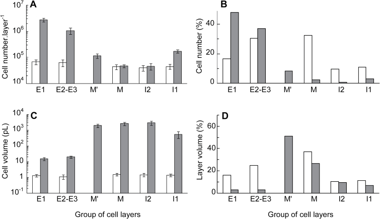
In angiosperms, the ovary wall resumes growth after pollination through a balanced combination of cell division and cell expansion. The quantitative pattern of these events remains poorly known in fleshy fruits such as tomato (Solanum spp.), in which dramatic growth of the pericarp occurs together with endoreduplication. Here, this pattern is reported at the level of each of the cell layers or groups of cell layers composing the pericarp, except for vascular bundles. Overall, cell division and cell expansion occurred at similar rates for 9 days post anthesis (DPA), with very specific patterns according to the layers. Subsequently, only cell expansion continued for up to 3-4 more weeks. New cell layers in the pericarp originated from periclinal cell divisions in the two sub-epidermal cell layers. The shortest doubling times for cell number and for cell volume were both detected early, at 4 DPA, in epicarp and mesocarp respectively, and were both found to be close to 14 h. Endoreduplication started before anthesis in pericarp and was stimulated at fruit set. It is proposed that cell division, endoreduplication, and cell expansion are triggered simultaneously in specific cell layers by the same signals issuing from pollination and fertilization, which contribute to the fastest relative fruit growth early after fruit set.

Keywords: Cell division; Solanum lycopersicum; cell expansion; endoreduplication; fruit; fruit set; growth rate; pericarp; tomato..


Figures

Fig. 1.
Fig. 1.
The structure of tomato pericarp during fruit growth. Tomato pericarp was observed on resin equatorial sections of ovaries at floral stages 11 (A) and anthesis (B), and of fruits at 4 DPA (C) and 36 DPA (D). Cell layers are named according to their relative positions: E1, outer epidermis; E2 and E3, two cell layers below E1; M, ~4 cell layers present at anthesis and surrounding vascular bundles (blue circles); I2, cell layer just close to I1, the inner epidermis. The M’ cell layers are formed after anthesis mostly from E2 and E3 but also, to a lesser, non-continuous extent from I2 (green asterisks, C, D). Yellow arrows show new cell walls indicative of recent periclinal cell divisions in E2 and I2 cell layers (A, B). Extracellular spaces at the border between M and adjacent layers E3, M’, or I2 are red. They are also present within M layers (not shown) but scarcely between the cells of other cell layers.
Fig. 2.
Fig. 2.
Fruit and pericarp growth at the time of fruit set. (A) Fruit volume and pericarp volume; (B) pericarp thickness and number of cell layers across the pericarp; (C) ploidy distribution within whole ovaries (up to 1 DPA) or dissected pericarp (2–4 DPA). Data are mean ± SD of measurements of 5.3 ± 1.8 ovaries or fruits in A and B, and of 4.0 ± 1.2 ovaries or fruits in C. The vertical dotted line marks the time of anthesis. The time scales for flower stages and for the post-anthesis period are not the same.
Fig. 3.
Fig. 3.
Mitotic activity in the tomato pericarp at fruit set. Mitoses were analysed in pericarp vibratome sections after DAPI staining at 1, 2, 3, and 4 DPA. Cell layers are named as in Fig. 1. A total number of 137 427 nuclei were analysed in the whole pericarp of 104 fruits, of which 30%, 19%, 36%, and 15% were located in E1, E2–E3, M’–M, and I2–I1 cell layers, respectively. (A) Mitotic indices (mean ± SE) including only metaphase, anaphase, and telophase figures were measured in each of the different cell layers of pericarp in seven fruits (1 and 2 DPA) or in 45 fruits (3 and 4 DPA). The horizontal line shows that there was no significant variation in mitotic index in a given cell layer within 1–4 DPA. (B) The orientation of cell division planes was determined from the pattern of mitotic nuclei as detailed in ‘Materials and methods’ (Supplementary Fig. S1B–F).
Fig. 4.
Fig. 4.
Time-course of cell number and cell volume in various cell layers of tomato pericarp during fruit growth. Each panel shows the evolution of cell number and of cell volume in one given cell layer. (A) E1 cell layer; (B) E2 cell layer; (C) M’ cell layer; (D) M cell layer; (E) I2 cell layer; (F) I1 cell layer. Variables are expressed as relative values with reference to the value of 1 at anthesis, indicated by the vertical dotted line. Two independent experiments are shown, first from anthesis to 36 DPA (dark lines and symbols), and second from flower bud stage 11 to 4 DPA (red lines and symbols). In current conditions, ~7 and 2 days separated stage 11 and stage 18 from anthesis, respectively. For the M’ cell layer (C), which results from periclinal cell divisions from E2 cells, the reference values are the values of the E2 cell layer at anthesis, and the variables are indicated from 4 and 2 DPA onwards in experiments 1 and 2, respectively. The data are mean ± SD for 3.5 ± 1.0 fruits in experiment 1 and 5.3 ± 1.8 fruits in experiment 2. The time scale is different for flower stages and DPA.
Fig. 5.
Fig. 5.
Cell multiplication and cell expansion during pericarp growth. The main panel shows data from experiment 1 in Fig. 4. The data show the mean ± SD values of pericarp volume, total cell number in pericarp, and cell volume according to fruit age. They are expressed as relative values with reference to the value of 1 at anthesis. The inset shows relative values ± SD of cell number and cell volume in experiment 2 from Fig. 4 for floral stages 11 up to 4 DPA. The following absolute values were found at anthesis in experiment 1 and 2, respectively: whole pericarp volume 0.539 ± 0.046 µL and 0.849 ± 0.311 µL; total cell number 0.411 ± 0.085 × 10⁶ and 0.562 ± 0.113 × 10⁶; mean cell volume 1.36 ± 0.30 pL and 1.49 ± 0.33 pL. This figure can be found in colour at JXB online.
Fig. 6.
Fig. 6.
Characteristics of the different groups of cell layers in tomato pericarp at the beginning and the end of fruit growth. The data are from experiment 1 in Fig. 4 and show values at 0 DPA (white bars) and at 36 DPA (grey bars). (A) Cell number in each single cell layer (mean ± SD). (B) Relative value of cell number in each group of cell layers with reference to the total pericarp cell number. (C) Mean cell volume ± SD in each group of cell layers. (D) Relative volume occupied by each group of cell layers in the pericarp. At anthesis and at 36 DPA, there was one cell layer in groups E1, I2, and I1, two cell layers in group E2–E3, and four cell layers in group M. There was 0 cell layers in group M’ at anthesis and 6.4 layers at 36 DPA.
Fig. 7.
Fig. 7.
Relative rates of fruit and pericarp growth, cell production, and cell volume increase in tomato. The data are from experiment 1 reported in Figs 4 and 5. Each point represents the slope of ln(variable) over three successive daily time points. (A) Relative rates of fruit and pericarp growth in volume. (B) Relative rates of cell production in pericarp and in E1 cell layer. (C) Relative rates of cell expansion in volume in pericarp and in the M cell layer.

References

    1. Barow M. 2006. Endopolyploidy in seed plants. Bioessays 28, 271–281. - PubMed
    1. Beemster GT, De Veylder L, Vercruysse S, et al. 2005. Genome-wide analysis of gene expression profiles associated with cell cycle transitions in growing organs of Arabidopsis. Plant Physiology 138, 734–743. - PMC - PubMed
    1. Bergervoet J, Verhoeven H, Gilissen LJW, Bino R. 1996. High amounts of nuclear DNA in tomato (Lycopersicon esculentum Mill.) pericarp. Plant Science 116, 141–145.
    1. Bertin N, Genard M, Fishman S. 2003a. A model for an early stage of tomato fruit development: cell multiplication and cessation of the cell proliferative activity. Annals of Botany 92, 65–72. - PMC - PubMed
    1. Bertin N, Borel C, Brunel B, Cheniclet C, Causse M. 2003b. Do genetic make-up and growth manipulation affect tomato fruit size by cell number, or cell size and DNA endoreduplication? Annals of Botany 92, 415–424. - PMC - PubMed

MeSH terms